IMAGE

Figure 3

ID
ZDB-IMAGE-230828-39
Source
Figures for Ha et al., 2023
Image
Figure Caption

Figure 3

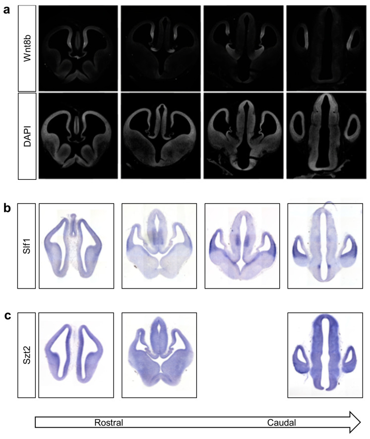
Wnt8b and Slf1 have spatially restricted expression patterns in the developing mouse brain. Coronal 20 μm frozen sections of mouse brain harvested at embryonic day 12. (a) Top row shows expression of Wnt8b measured using indirect immunofluorescent staining. Detection by rabbit anti-WNT8B primary antibody. Nuclei in the same section visualised using DAPI staining are shown in the second row to define morphology of the section. (b) Detection of Slf1 transcripts by in situ hybridisation using an in vitro transcribed anti-sense riboprobe. (c) Detection of Szt2 transcripts by in situ hybridisation using an in vitro transcribed anti-sense riboprobe.

Acknowledgments
This image is the copyrighted work of the attributed author or publisher, and ZFIN has permission only to display this image to its users. Additional permissions should be obtained from the applicable author or publisher of the image. Full text @ Genes (Basel)