IMAGE

Figure 9

ID
ZDB-IMAGE-230712-22
Source
Figures for Gora et al., 2023
Image
Figure Caption

Figure 9

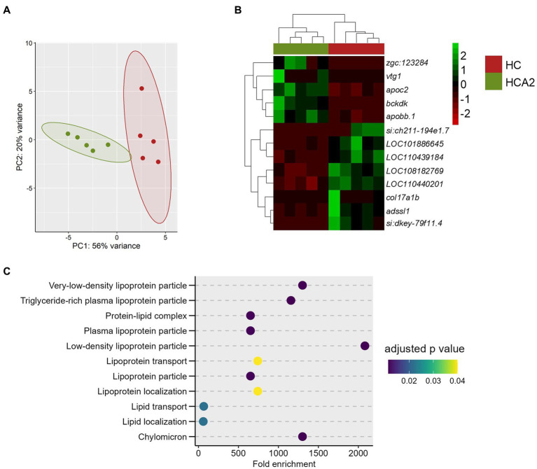
Differences in the transcriptome of the HCA2 and HC groups. Principal component analysis (A) and Hierarchical clustering (B) of the differentially expressed genes (DEGs) in the HCA2 group compared to the HC group. Transcripts with an adjusted p value (Benjamini-Hochberg method) < 0.05 and |Log2 fold change| ≥ 1 were considered significantly differentially expressed. Dot-plot (C) showing the enriched GO terms, by considering the upregulated genes in the HCA2 vs. HC group comparison. Each treatment group consisted of at least five biological replicates.

Acknowledgments
This image is the copyrighted work of the attributed author or publisher, and ZFIN has permission only to display this image to its users. Additional permissions should be obtained from the applicable author or publisher of the image. Full text @ Front Nutr