
|
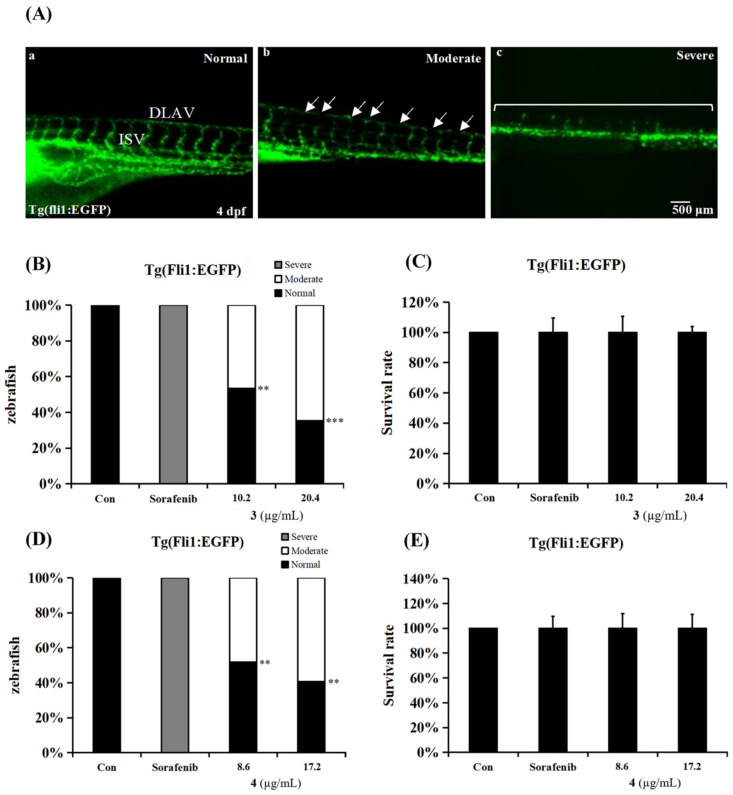
Fig. 5
Penisterine C (3) and penisterine D (4) exert anti-angiogenic capacities in vivo. (A) Representative images of the trunk vasculature in Tg (fli1:EGFP) zebrafish embryos incubated with compounds or sorafenib at 4 days post-fertilization (dpf). Arrows indicate impaired at intersegmental vessel (ISV) and dorsal longitudinal anastomic vessel (DLAV). (B) Quantitative analysis presents percentage of fish embryos incubated with 3 with defective vasculature. ** p < 0.01, and *** p < 0.001 compared with the control group. (C) Quantitative analysis presents percentage of survival rate of fish embryos incubated with 3. (D) Quantitative analysis presents percentage of fish embryos incubated with 4 with defective vasculature. ** p < 0.01 compared with the control group. (E) Quantitative analysis presents percentage of survival rate of fish embryos incubated with 4. The zebrafish treated with 1% DMSO was used as a negative control, and 10 µg/mL sorafenib was used as a positive control.