
|
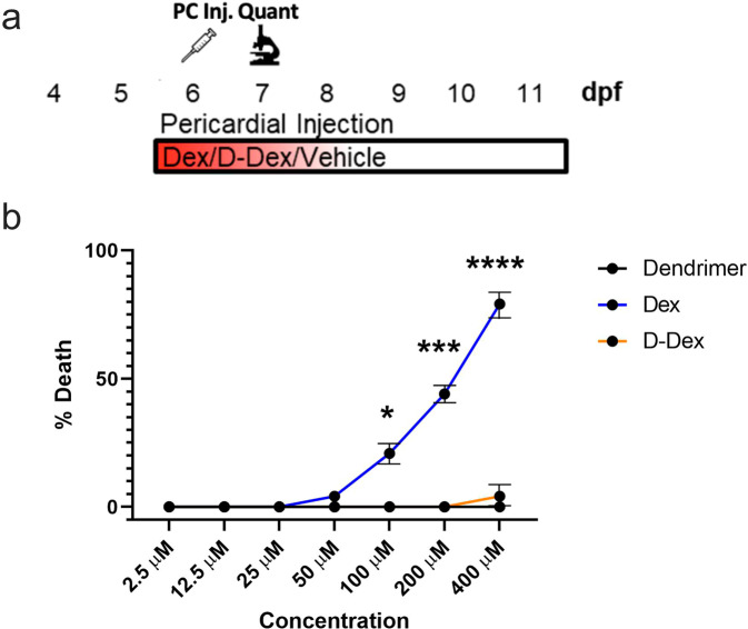
Fig. 2
a Schematic of injection assay to test dendrimer, Dex and D-Dex toxicity. At 6 dpf, dendrimer, “free” Dex, or D-Dex were injected into the pericardium of zebrafish larvae (~10 nL injected volume, 2.5-400 µM for Dex and D-Dex). At 7 dpf, toxicity was quantified based on percent survival across 24 fish per condition over two trials. b Line graph indicating average toxicity at 7 dpf for dendrimer (black line), Dex (blue line), and D-Dex (orange line). Comparisons between Dex or D-Dex and dendrimer alone controls showed statistically significant increases in toxicity for Dex-injected larvae only (100-400 μM, *p ≤ 0.05, ***p ≤ 0.001, ****p ≤ 0.0001, respectively).