
|
Fig. 1
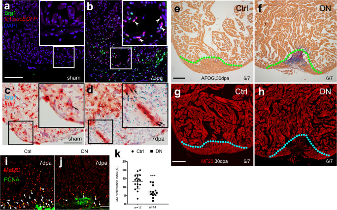
a, b Immunofluorescence staining of Brg1 and EGFP on paraffin sections of Tg(fli1:nucEGFP) transgenic hearts from sham-operated (a) and injured zebrafish hearts (b) at 7 dpa (arrowheads, Brg1- and EGFP-positive endothelial cell nuclei). c, d RNAscope in situ hybridization of brg1 and kdrl probes in frozen sections from sham-operated (c) and injured hearts (d) at 3 dpa (arrows, brg1- and kdrl-positive endothelial cells). The upper right corner presented in (a–d) is a high-magnification image of the framed area. e–h Representative images of Acid fuchsin orange G (AFOG) staining (e, f) and immunofluorescence with anti-myosin heavy chain (MF20) (g, h) of heart sections from control siblings Tg(ubi:LoxP-DsRed-STOP-LoxP-dn-xbrg1) (Ctrl) and endothelium-specific dominant-negative