
|
Fig. 6
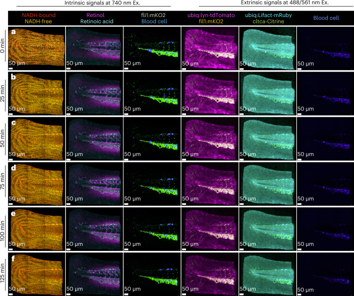
HyU’s increased sensitivity provides a simple solution for the challenging task of imaging timelapse data at six time points (125 min) for both intrinsic signals and extrinsic signals of a quadra-transgenic zebrafish: Gt(cltca-citrine);Tg(ubiq:lyn-tdTomato;ubiq:Lifeact-mRuby;fli1:mKO2). a–f, Volumetric renderings of HyU results for time points acquired at 25 min intervals reveal the high-contrast and high-multiplexed labels of NADH-bound (red), NADH-free (yellow), retinoid (magenta), retinoic acid (cyan), mKO2 (green) and AF from blood cells (blue) when excited at 740 nm. Further extrinsic signals for mKO2 (yellow), tdTomato (magenta), mRuby (cyan), citrine (green) and blood cells AF (blue) are also readily unmixed using HyU when exciting the sample at 488/561 nm. HyU provides the capacity to simultaneously multiplex nine signals in a live sample over long periods of time, a previously unexplored task. Scale bars, 50 µm. The sample depicted is representative of 28 experimental sessions each with three to five biological replicates, yielding similar results.