
|
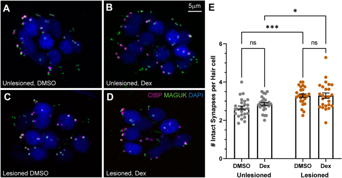
Fig. 4
Dexamethasone exposure did not alter the number of afferent synapses in neuromast hair cells. A–D Representative maximum intensity projection images of 7 dpf pLL neuromasts, with hair cell presynaptic ribbons labeled via CtBP antibody (magenta), post-synaptic densities labeled via MAGUK antibody (green), and hair cell nuclei labeled with DAPI (blue). Neuromasts were either unlesioned (A, B) or lesioned with CuSO4 (C, D), then allowed to regenerate for 48 h while exposed to DMSO (carrier) (A, C) or dexamethasone (B, D). E Scatter plots with mean and SEM bars showing no significant difference in the number of intact synapses in dexamethasone-treated fish, compared to DMSO controls for both lesioned and intact neuromasts. Each dot represents a single neuromast on an individual fish. Notably, a significantly greater number of intact synapses per hair cell were observed in regenerating neuromasts relative to neuromasts that were not lesioned (adjusted ***P < 0.001 (DMSO), *P < 0.05 (Dex)). N = 5–10 fish per condition per trial; 4 experimental trials