
Image
|
Figure Caption
Figure 7
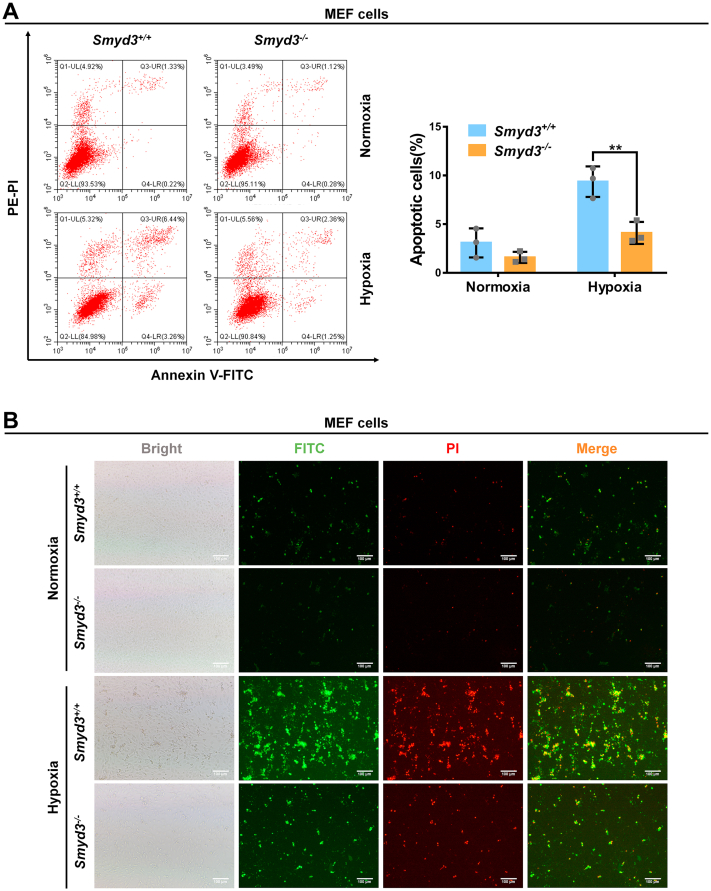
Disruption of SMYD3 protects cells against hypoxia-induced apoptosis. A, apoptotic cells in Smyd3-deficient or wildtype MEF cells (Smyd3−/− or Smyd3+/+) under normoxia or hypoxia detected by flow cytometry analysis. Data show mean ± SD; Student’s two-tailed t test. ∗∗p < 0.01. Data from three independent experiments. B, apoptotic cells in Smyd3-deficient or wildtype MEF cells (Smyd3−/− or Smyd3+/+) under normoxia or hypoxia detected by fluorescence microscopy. Scale bar = 100 μm. MEF, mouse embryonic fibroblast.
Acknowledgments
This image is the copyrighted work of the attributed author or publisher, and
ZFIN has permission only to display this image to its users.
Additional permissions should be obtained from the applicable author or publisher of the image.
Full text @ J. Biol. Chem.