
Image
|
Figure Caption
Figure 4
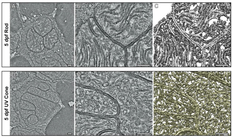
Tomograms show the compact association of mitochondria within the inner segments of UV cone and rod photoreceptors at 5 dpf. (A–C) Tomography data of rod and (D,E) UV cone inner segment mitochondria. (A,D) Single slices from the tomograms with (B,E) higher magnification views. (C–F) Surface rendering of the tomography data allows the architecture of the mitochondria to be assessed in 3D. The rod has elongated and tightly associated cristae membranes, whereas the UV cone has cristae that are wider. Scale bars = 250 nm.
Acknowledgments
This image is the copyrighted work of the attributed author or publisher, and
ZFIN has permission only to display this image to its users.
Additional permissions should be obtained from the applicable author or publisher of the image.
Full text @ Cells