
Image
|
Figure Caption
Fig. 1
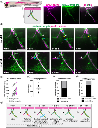
Perineurial glia form a bridge and phagocytose debris after spinal motor nerve injury. (a) (Left) Diagrammatic representation of a 6 dpf zebrafish larva with a single peripheral spinal motor nerve (inset) displayed. Motor axon (magenta), ensheathed Schwann cells (orange), perineurial glia (green). (Right) Representative images of an in vivo 6 dpf spinal motor nerve, where olig2 labels motor axons (magenta) and nkx2.2a labels perineurial glia (green). (b–f) n = 11 nerves in 5 larvae. (b) Representative stills taken from time-lapse movies of perineurial glia (green) and motor axons (magenta) in 5 or 6 dpf larvae injured on either the caudal or rostral side of the spinal motor nerve. Stills are shown in the first 6 h post injury (hpi). The dashed circle indicates the injury site. Blue arrows denote phagocytic vesicles in perineurial glia. White arrows follow the proximal end and yellow arrows follow the distal end of the perineurial glial bridge. (c) Quantification of perineurial glial (PG) bridging timing. Timing of bridging initiation (green) and completion (magenta) was recorded in minutes post injury. (d) Quantification of perineurial glial (PG) bridging duration in minutes (mean: 150.54 ± 18.25 min). (e) Quantification of the type of perineurial glial (PG) bridging observed. Proximal and distal bridging (45% of nerves, gray) or proximal only bridging (55% of nerves, black). (f) Quantification of perineurial glial (PG) phagocytosis after injury. Phagocytic vesicles on both proximal and distal perineurial glial stumps (27% of nerves, gray) or only on proximal perineurial glial stumps (73% of nerves, black). (g) Representative timeline of the first 48 h following spinal motor nerve injury in 5 to 6 dpf zebrafish. Motor axon (magenta), Schwann cells (orange), perineurial glia (green), macrophages (blue). Scale bars, 25 μm.
Acknowledgments
This image is the copyrighted work of the attributed author or publisher, and
ZFIN has permission only to display this image to its users.
Additional permissions should be obtained from the applicable author or publisher of the image.
Full text @ Glia