
|
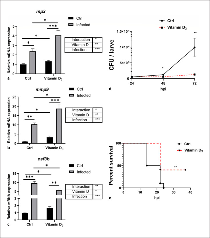
Fig. 5
VD3 restrains pathogen infection in zebrafish. a–c Transcript levels of mpx (a), mmp9 (b), and csf3b (c) in unstimulated and E. tarda-exposed zebrafish larvae, pretreated with 0 or 100 nM VD3 (n = 4 replicates/group, 10–20 larvae/replicate). Two-way ANOVA with Sidak test was used to test significance. d Zebrafish larvae at 3 dpf were immersed with 1 × 10⁸ CFU/mL E. tarda. After 24, 48, and 72 h, the bacteria burden in the larvae was counted (n ≥ 24 larvae/group/experiment, 3 independent experiments). e Zebrafish larvae at 3 dpf were pretreated with control buffer or VD3 (50 nM) for 48 h. Afterward, the larvae were microinjected with E. tarda (approximately 200 bacteria/larva). The survival of the larvae in each group was recorded up to 36 hpi (n = 10 larvae/group/experiment, 3 independent experiments). Statistical analysis was conducted by Log-rank test. Data are represented as mean ± SEM. *p < 0.05, **p < 0.01, ***p < 0.001. csf3b, colony-stimulating factor 3b; mmp9, matrix metalloproteinase9; dpf, days post fertilization; VD3, vitamin D3; E. tarda, Edwardsiella tarda; hpi, hours post infection; mpx, myeloperoxidase.