
|
Fig. 3
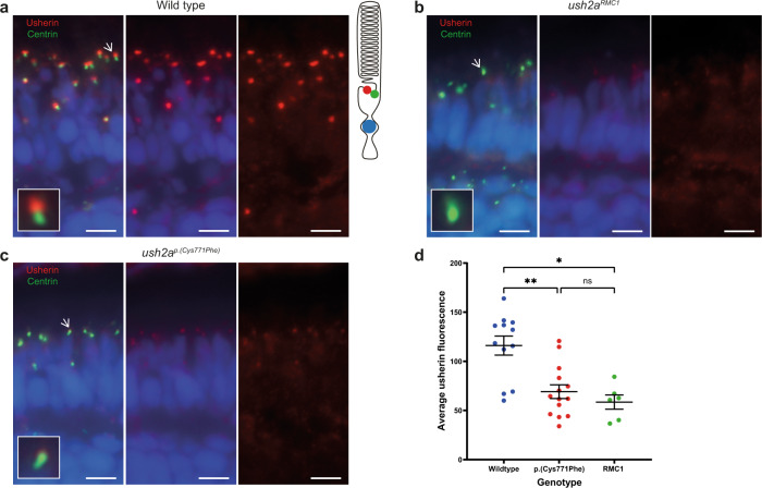
a In wildtype zebrafish larval eyes (n = 12 eyes), usherin (red signal) localizes at the photoreceptor periciliary membrane adjacent to the basal body and connecting cilium marker centrin (green signal) as shown by the schematic representation of a photoreceptor on the right. A magnification of one photoreceptor (indicated by an arrow) is depicted in the inlay. b In ush2armc1 knock-out larvae usherin is not detectable (n = 6 eyes). c Localization of usherin at the photoreceptor periciliary membrane was strongly reduced in eyes of ush2ap.(Cys771Phe) larvae (n = 14 eyes) as compared to wildtype. d A Kruskal–Wallis test was performed based on the average of the mean grey value for usherin adjacent to each centrin spot and confirmed a significant decrease of usherin localization adjacent to centrin for the usherinp.(Cys771Phe) and the usherinRMC1 models. The average grey value per retinal section was plotted in a scatter plot (mean ± SEM). Nuclei are stained with DAPI (blue signal). Scale bar: 5 µM. **p: 0.0094, *p: 0.0107, ns not significant.