
|
Fig. 2
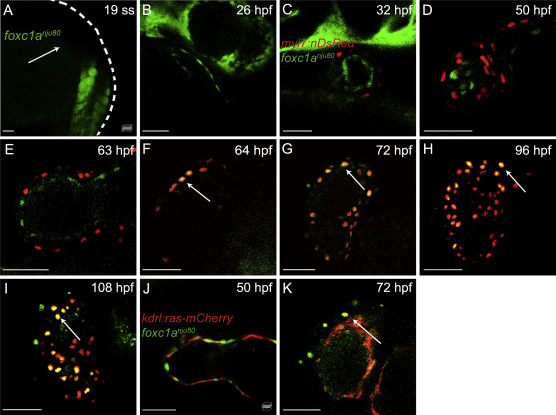
Fig. 2. Foxc1a is expressed in ventricular cardiomyocytes after 64 hpf. A and B: Confocal sagittal sections of foxc1anju80 embryos at 19 ss and 26 hpf. The white arrow in (A) points to the cardiac cone region. A lateral view with the abdomen oriented to the left and the head upward (A). A lateral view with the abdomen oriented to the right and the head upward (B). C–I: Confocal sagittal sections of Tg(myl7:nDsRed);foxc1anju80/+ in-crossed embryos from 32 hpf to 108 hpf. All images are ventral views with the heads oriented upward. The myl7+ cells are labeled in red, and the foxc1a+ cells are labeled in green. The white arrows in (F–I) indicate representative colocalized cells. J and K: Representative projections of in vivo confocal images show the representative hearts of Tg(kdrl:ras-mCherry);foxc1anju80/+ in-crossed embryos. Both images show ventral views with the head upward. The white arrow in (K) indicates a representative foxc1a+ cell. The kdrl+ cells are labeled in red, and the foxc1a+ cells are labeled in green. Scale bars, 50 μm. ss, somite stage; hpf, postfertilization.
Reprinted from Journal of genetics and genomics = Yi chuan xue bao, 49(6), He, L., Zhang, Q., Jiang, D., Zhang, Y., Wei, Y., Yang, Y., Li, N., Wang, S., Yue, Y., Zhao, Q., Zebrafish foxc1a controls ventricular chamber maturation by directly regulating wwtr1 and nkx2.5 expression, 559-568, Copyright (2021) with permission from Elsevier. Full text @ J. Genet. Genomics