IMAGE

Figure 6

ID
ZDB-IMAGE-220625-7
Source
Figures for Shi et al., 2022
Image
Figure Caption

Figure 6

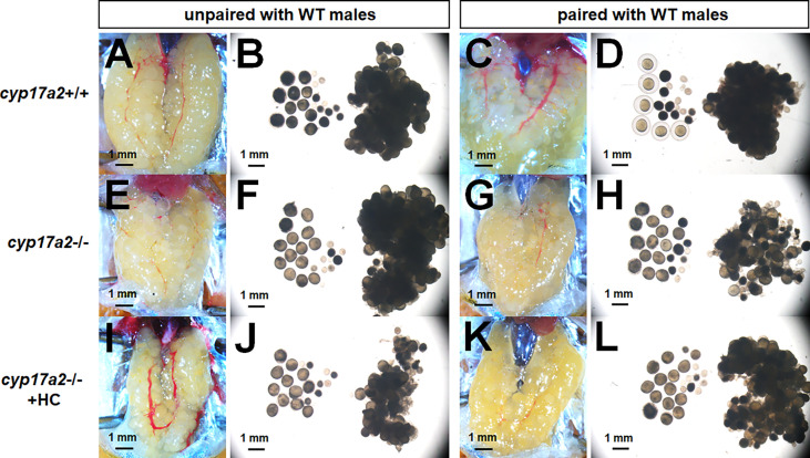
The over-activated oocytes in cyp17a2-/- females. (A–D) Under visual microscopy, the oocytes from control females unpaired and paired with WT males were not transparent and transparent, respectively (n = 5). (E–H) The oocytes from cyp17a2-/- females unpaired and paired with WT males were both transparent (n = 5). (I–L) The 20 nM hydrocortisone treatment in cyp17a2-/- females did not promotes oocyte maturation as observed under visual microscopy (n = 5).

Acknowledgments
This image is the copyrighted work of the attributed author or publisher, and ZFIN has permission only to display this image to its users. Additional permissions should be obtained from the applicable author or publisher of the image. Full text @ Front Endocrinol (Lausanne)