
Image
|
Figure Caption
Fig. 4
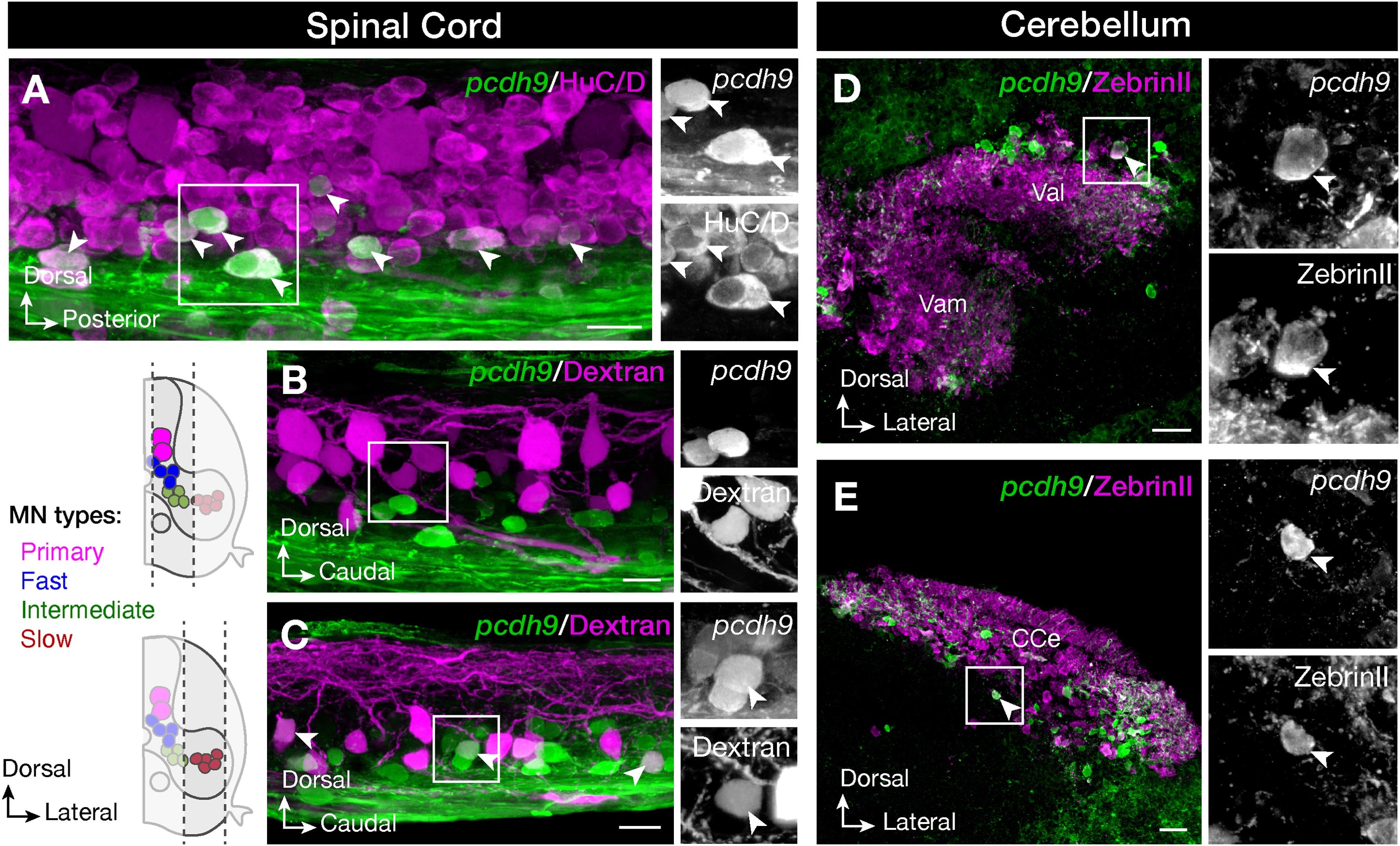
Tg(pcdh9:hs:GFP) juvenile zebrafish exhibit expression both in the spinal cord and in the cerebellum. All the pcdh9 positive cells expressed the pan-neuronal marker HuC/D (A). Retrograde labelling of motor neurons showed a specific expression of pcdh9 in a portion of slow secondary motor neurons but not in primary, or fast and intermediate secondary motor neurons (B, C). In the cerebellum, pcdh9 positive cells were positive for ZebrinII both in the valvula lateralis (Val) and valvula medialis (Vam) (D) and in the corpus cerebelli (CCe) (E). Scale bars equal 20 μm.
Figure Data
Acknowledgments
This image is the copyrighted work of the attributed author or publisher, and
ZFIN has permission only to display this image to its users.
Additional permissions should be obtained from the applicable author or publisher of the image.
Reprinted from Gene expression patterns : GEP, 44, Habicher, J., Manuel, R., Pedroni, A., Ferebee, C., Ampatzis, K., Boije, H., A new transgenic reporter line reveals expression of protocadherin 9 at a cellular level within the zebrafish central nervous system, 119246, Copyright (2022) with permission from Elsevier. Full text @ Gene Expr. Patterns