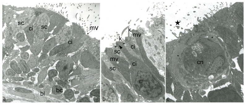
IMAGE

Figure 1

ID
ZDB-IMAGE-220518-104
Source
Figures for Aragona et al., 2022
Image
Figure Caption

Figure 1

Transmission electron micrographs of ultrathin sections showing the surface of olfactory epithelium adult zebrafish: (a) ci, ONR ciliated; mv, ONR microvillous; sc, supporting cells; bc, basal cells; bl, basal lamina. (b) ci, ONR ciliated; n,“checkerboard” nucleus of ciONR; K, kinocilia; mv, ONR microvillous; sc, supporting cells; kinocilium basal body foot (arrowhead), junctional complex with neighboring supporting cells (arrows). (c) cn, crypt neurons with several cilia within the crypt (star); special supporting cells (*).

Acknowledgments
This image is the copyrighted work of the attributed author or publisher, and ZFIN has permission only to display this image to its users. Additional permissions should be obtained from the applicable author or publisher of the image. Full text @ Int. J. Mol. Sci.