
|
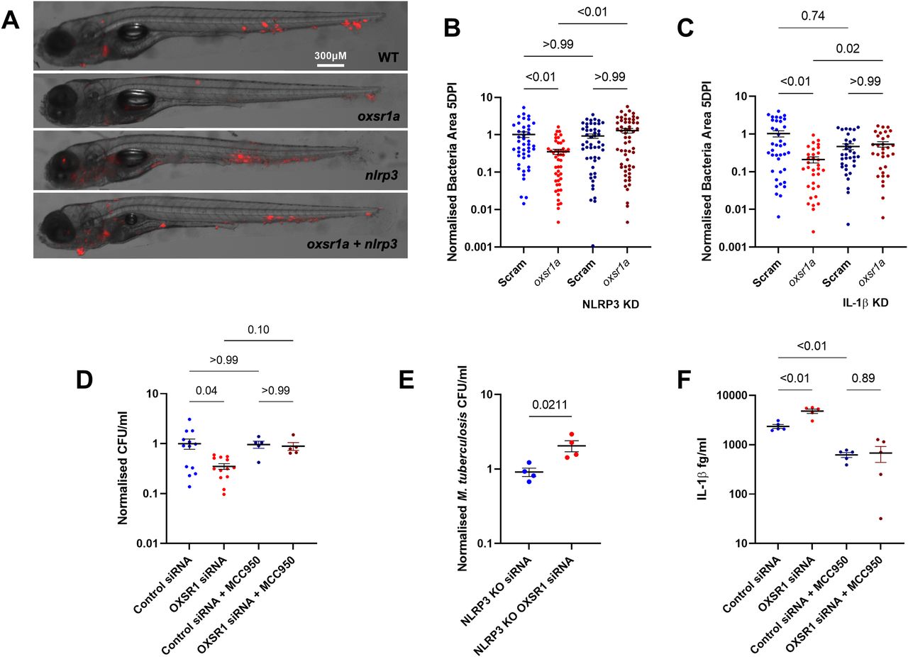
Fig. 4
(A) Representative images of M. marinum–tdTomato (red) bacterial burden in 5 dpi WT, mosaic F0 oxsr1a, nlrp3, and dual oxsr1a nlrp3 crispant embryos. (B) Quantification of WT M. marinum bacterial burden in WT, mosaic F0 oxsr1a, nlrp3, and dual oxsr1a nlrp3 crispant embryos. Combined results of three biological replicates. (C) Quantification of WT M. marinum bacterial burden in WT, mosaic F0 oxsr1a, Il1b, and dual oxsr1a Il1b crispant embryos. Combined results of two biological replicates. (D) Quantification of intracellular M. tuberculosis bacterial burden in WT and OXSR1 knockdown differentiated THP-1 cells at 3 dpi. (E) Quantification of intracellular M. tuberculosis burden in WT and OXSR1 knockdown differentiated NLRP3 knockout THP-1 cells at 3 dpi. (F) IL-1β content in the supernatant of M. tuberculosis–infected WT and OXSR1 knockdown differentiated THP-1 cells at 3 dpi. For cell experiments (D, E, F): each dot represents the CFU from an infected well in a single representative experiment and the experiment was repeated three times.