
Fig. 4
- ID
- ZDB-IMAGE-220512-16
- Publication
- Xue et al., 2019 - Novel cathepsin K inhibitors block osteoclasts in vitro and increase spinal bone density in zebrafish
- All Figures
- Figures for Xue et al., 2019
|
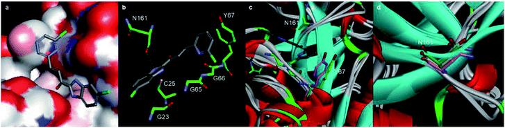
Fig. 4 Molecular docking calculations of the binding mode of A22 with Cat K (PDB code: 4dmy). (a) and (b) The calculated interactions of A22 (grey) with amino acids (green) in the active pocket of Cat K. (c) Overlay of the crystal structures of Cat L (PDB code: 6ezp), Cat S (PDB code: 4bs5) and Cat B (PDB code: 6ay2) with Cat K. N161 (green) of Cat K located at a loop region where the conformations of the Cat L, S, B and K are different. Compared to the orientation of Y193 (purple) of Cat S, Y67 (green) of Cat K has much less clash with pyridine ring of A22. (d) Backbone carbonyl groups of N161 of Cat K, N286 (purple) of Cat S and D162 (yellow) of Cat L.