
|
Figure 5
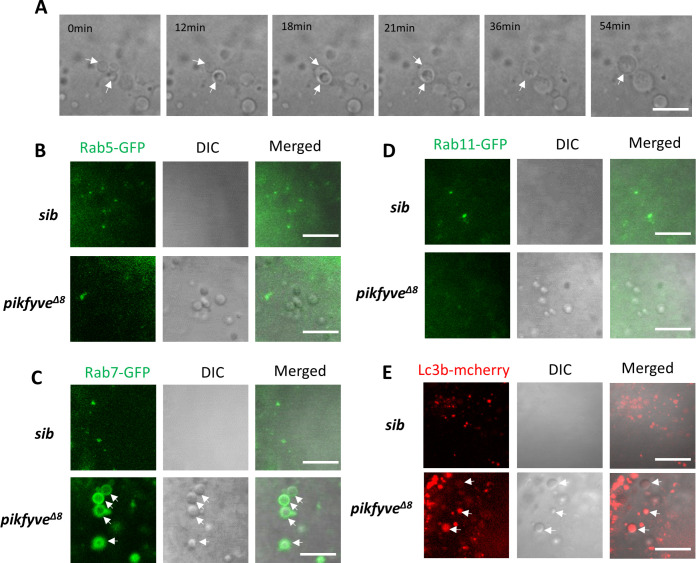
(A) Time-lapse imaging indicating the dynamic changes of vacuole formation in the lens of 4-dpf pikfyveΔ8 mutants. White arrows indicate the fusion process of two small vacuoles. (B) Representative images showing the lens of 3.5-dpf siblings and pikfyveΔ8 mutants injected with gfp-rab5c mRNA. (C) Representative images showing lens of 3.5-dpf siblings and pikfyveΔ8 mutants injected with gfp-rab7 mRNA. (D) Representative images showing lens of 3.5-dpf siblings and pikfyveΔ8 mutants injected with gfp-rab11a mRNA. (E) Representative images showing lens of 3.5-dpf siblings and pikfyveΔ8 mutants injected with mcherry-lc3b mRNA. All experiments were repeated three times. All the scale bars represent 10 μm.Characterization of vacuoles in pikfyveΔ8 mutants.