
|
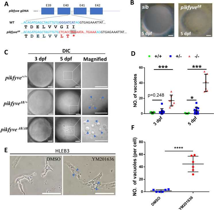
Figure 3
(A) A schematic diagram showing the generated pikfyveΔ8 mutant allele. The underlined base pairs are the sgRNA target. The deleted base pairs are shown in dark blue while inserted ones are shown in red. The stop codon introduced in the mutant form is shown in the grey box. (B) Representative images showing the lens of sibling and pikfyveΔ8 mutants at 5 dpf. (C) Representative differential interference contrast (DIC) images showing the lens of pikfyve+/+, pikfyve+/Δ8, and pikfyveΔ8/Δ8 embryos at 3 dpf and 5 dpf. The scale bars represent 10 μm in (B) and (C). (D) Quantification of vacuole number in the lens of pikfyve+/+, pikfyve+/Δ8, and pikfyveΔ8/Δ8 embryos at 3 dpf (n=7 for pikfyve+/+; n=10 for pikfyve+/Δ8; n=7 for pikfyveΔ8/Δ8) and 5 dpf (n=7 for pikfyve+/+; n=11 for pikfyve+/Δ8; n=6 for pikfyveΔ8/Δ8). (E) Representative images of HLEB3 cells treated with DMSO or PIKFYVE inhibitor YM201636 for 4 hr. The scale bars represent 25 μm. (F) Quantification of the vacuole numbers in (E). ****, p<0.0001, Student’s t-test. All experiments were repeated three times. See Figure 3—source data 1 for details.
Disruption of the PIPK domain of Pikfyve in zebrafish caused early-onset cataract.