
|
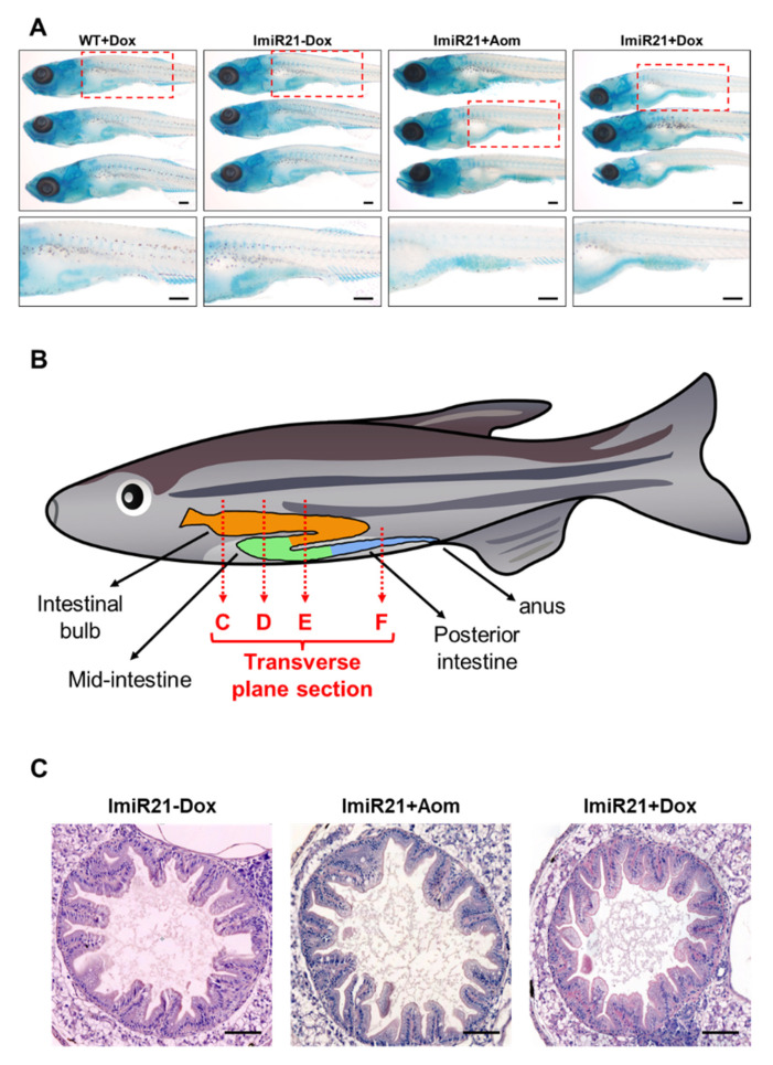
Figure 4
Characterization of inflammatory bowel disease (IBD)-like phenotype in ImiR-21. (A) Representative images of whole-mount alcian blue staining of WT + Dox, ImiR-21 ± Dox, and ImiR-21 + AOM at 21 days post-fertilization (dpf). Blue signals are the stained goblet cells in the injured villus of the posterior intestine of ImiR-21 + AOM and ImiR-21 + Dox animals under inflammatory conditions. Scale bar: 200 μm. (B) Schematic diagram of the intestinal structure of zebrafish. H&E-stained transverse plane sections in different parts of ImiR-21 ± Dox and ImiR-21 + AOM intestines (21 dpf) (intestinal bulb, mid-intestine, and posterior intestine are corresponded to panels (C–F)). Goblet cell hyperplasia is indicated by arrows. Scale bar: 50 μm.