
|
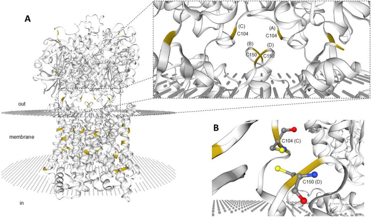
Fig. 1
(A) Three-dimensional appearance (lateral view) of the zebrafish Slc7a9(b0,+AT)-Slc3a1(rBAT) complex bound to arginine in the hetero-2–2-mer (i.e., heterotetramer made of 2 heterodimers) form and (B) snapshot of the putative disulfide bridge linking covalently one of the two heterodimers (i.e., the heterodimer made of chain C, Slc3a1 and chain D, Slc7a9). The model was built using Protein Data Bank Acc. No. 6li9.1 as a template. Cysteine residues only are highlighted (yellow). A, chain A; B, chain B; C, chain C; D, chain D. Chains A and C refer to zebrafish Slc3a1; chains B and D refer to zebrafish Slc7a9. C104, cysteine residue C104 on chain C, Slc3a1; C150, cysteine residue C150 on chain D, Slc7a9