
|
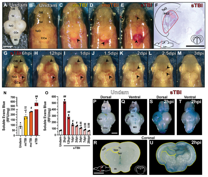
Figure 2
MMWD produces graded hematomas and blood–brain barrier disruption. (A) Isolated, undamaged whole brain with the major lobes labeled. Dorsal views of roya9;mitfaw2 (casper) undamaged (B) and TBI fish displaying vascular injury 4 hpi (C–E). Compared to undamaged controls, vascular injury resulted in hemorrhaging (arrowheads) in all severity levels that increased in a severity-dependent manner. (F) Coronal section of sTBI brain with intracerebral hematoma (red boundary). (G–M) Repeated dorsal view of an individual sTBI albinob4 fish across time, in which hemorrhaging (arrowheads) qualitatively peaked at 12 hpi, and gradually resolved by 3 dpi. (N) Following injury, a significant increase in solubilized Evans Blue dye represented disruption of the BBB in a severity-dependent manner. (O) Following sTBI, a statistically significant increase in solubilized Evans Blue dye occurred by 2 hpi and then gradually decreased until it reached control levels at 3 dpi (3 pooled brains/group, n = 2–3 groups). (P–U) Dorsal, ventral, and coronal views of isolated undamaged (P,Q,R) and sTBI brains at 2 hpi (S,T,U) from fish injected with Evans Blue dye as a qualitative measure of BBB integrity. Solid lines in (F,R,U) denote tissue boundaries, while dotted lines denote internal anatomical boundaries. Corpus cerebelli, CCe, hypothalamus, Ht, medial valvula cerebelli, Vam, medulla oblongata, MO, medulla spinalis, MS, olfactory bulb, OB, optic tectum, TeO, periventricular grey zone, PGZ, tectal ventrical, TeV, telencephalon, Tel, torus longitudinalis, TL, ventral optic tectum, VTeO, ventral telencephalon, VTel. Scale bars, (A–E,G–M,P–T) = 500 µm, (F,R–U) = 250 µm. Mean ± SEM is depicted in (N,O). Statistical analyses were performed with a One-way ANOVA followed by a Tukey post-hoc test. ## p < 0.01.