IMAGE

FIGURE 4

ID
ZDB-IMAGE-210707-9
Source
Figures for Li et al., 2021
Image
Figure Caption

FIGURE 4

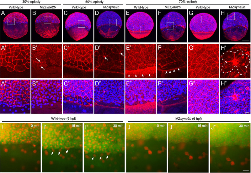
Disruption of Syne2b function affects F-actin organization, EVL cell shape and YSN movements. Phalloidin and DAPI staining of stage-matched embryos. (A–A″) Wild-type embryos at 30% epiboly show strong phalloidin staining in the cortex of EVL cells. A ring of YSN are apparent at the EVL margin. (B–B″) MZsyne2b embryos at 30% epiboly show reduced and clustered (arrows) phalloidin staining in EVL cells. Few YSN are present at the EVL margin. (C–C″) In wild-type embryos at 50% epiboly, phalloidin staining is present strongly in the cortex of EVL cells and uniformly in the yolk cell. YSN are still present at the EVL margin. (D–D″) In MZsyne2b embryos at 50% epiboly, phalloidin staining is clustered at multiple cell contact regions in the blastoderm (arrows) and is disrupted in the yolk cell. Few YSN are scattered in the yolk cell. (E–E″) In wild-type embryos at 70% epiboly, thick actin rings are formed around the blastoderm margin (arrowheads). (F–F″) MZsyne2b embryos at 70% epiboly form weak and thin marginal actin rings (arrowheads). YSN remain scattered in the yolk cell. (G–G″) Regular cortical F-actin and cell shape in the blastoderm of wild-type embryos. (H–H″) Severely disrupted cell shape and rearrangements in the blastoderm of MZsyne2b embryos, with the occurrence of rosette structures (broken lines). (I,J″) Still frames from time-lapse imaging show YSN movements at 50% epiboly. Note that YSN emerge from the front of the EVL margin during epiboly in wild-type embryos (arrows). Scale bars: (A–H) 200 μm; (A′–H″) 20 μm; (I–J″) 100 μm.

Figure Data
Acknowledgments
This image is the copyrighted work of the attributed author or publisher, and ZFIN has permission only to display this image to its users. Additional permissions should be obtained from the applicable author or publisher of the image. Full text @ Front Cell Dev Biol