
|
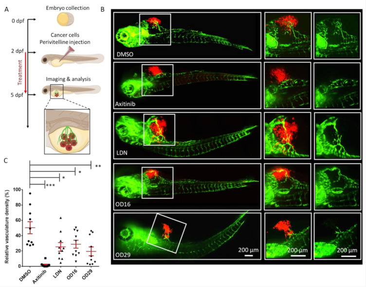
Figure 7 OD16 and OD29 inhibit vessel invasion in a breast cancer xenograft model in zebrafish. (A) Schematic representation of the experimental procedure. Two dpf zebrafish embryos were injected with mCherry (red) labeled human MDA-MB-231 breast cancer cells in the perivitelline space. The injected zebrafish embryos were challenged with testing compounds in egg water for 3 days, and the newly formed vessels within the perivitelline were analyzed. (B) Representative images of the eGFP expressing blood vessels (green) and mCherry MDA-MB-231 cells (red) after treatment of the zebrafish embryos with vehicle control (DMSO), Axitinib, LDN-193189, OD16, or OD29 (0.5 µM) for 3 days. The medium (vessels and tumor cells are visualized) and right panels (only vessels are visualized) represent high magnification views of the area, indicated by white rectangles in the overviews (left panel) images. Scale bars represent 200 µm. (C) Quantification of the relative vasculature density induced by the injected MDA-MB-231 cells within the perivitelline space of the zebrafishes in each group. * p < 0.05, ** p < 0.005, *** p < 0.001.