
Image
|
Figure Caption
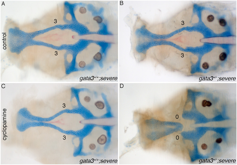
Fig 9 (A-D) Flat mounts of 5dpf gata3 b1075/b1075 neurocrania anterior to the left. (A-B) control untreated-embryos from the gata3 “severe” background showing normal stacking and fusion of trabeculae. Both of (A) wild type and (B) heterozygotes develop normally. (C-D) Cyclopamine-treated embryos. (C) Wild type embryo showing normal stacking and fusion of trabeculae (n = 22/22). (D) Heterozygote embryo displaying complete loss of trabeculae with severe clefts (n = 6/61).
Figure Data
Acknowledgments
This image is the copyrighted work of the attributed author or publisher, and
ZFIN has permission only to display this image to its users.
Additional permissions should be obtained from the applicable author or publisher of the image.
Full text @ PLoS Genet.