
|
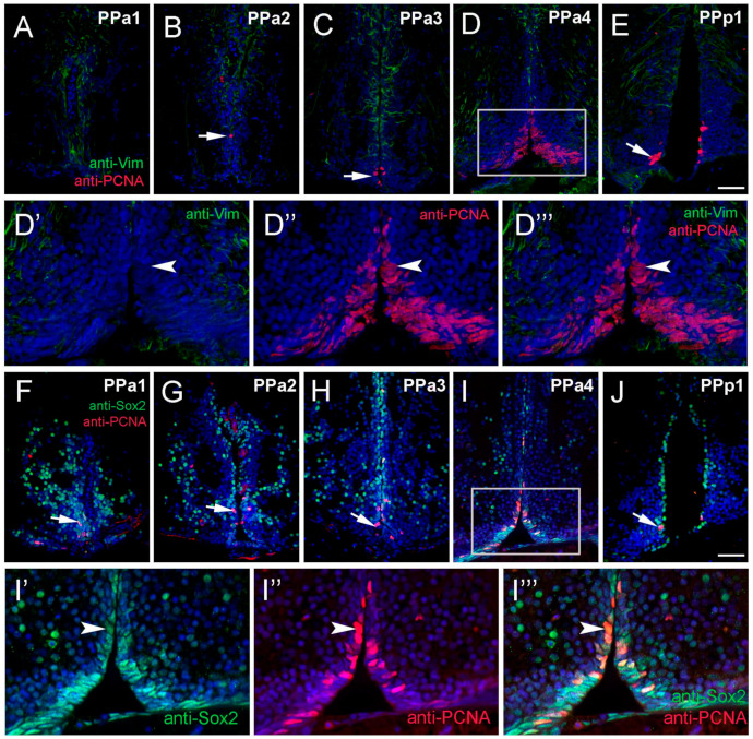
Figure 4 Proliferating cell nuclear antigen (PCNA) is located primarily in the PPa4/PPp1 transition region. (A–J) Transverse paraffin-sections of 5 µm of POA. (A–E) Vim+ cells (green) are localized to the dorsal region of the POA, in contrast, PCNA+ cells (red, arrow (B,C,E)) are found in the ventral of the POA. Vim+ labeling ((D’,D’’’), green) does not co-localize with anti-PCNA labeling ((D’’,D’’’), red). (F–H) Many Sox2+ cells (green) were observed in PPa1-3 and PPp1 (J) with few PCNA+ cells (red, arrows). (I) Sox2+ cells (green) and PCNA+ (red) in the ventral PPa4 region of the POA (boxed area) where the signals were co-localized in some cells ((I’–I’’’), arrowheads). All sections were labeled with DAPI (blue). Scale bar: 30 μm.