IMAGE

Fig. 3

ID
ZDB-IMAGE-210611-42
Source
Figures for Quint et al., 2021
Image
Figure Caption

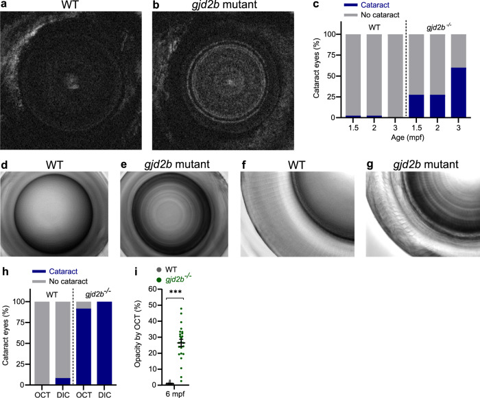
Fig. 3 Nuclear cataract in <italic>gjd2b</italic> (Cx35.1) mutants.

a, b Coronal SD-OCT sections of typical 3 mpf lenses of WT control (a) and gjd2b (Cx35.1) mutant (b) fish. c Proportion of cataractous lenses in 1.5 mpf, 2 mpf, and 3 mpf SD-OCT data (n = 40 eyes) indicating an increasing prevalence in gjd2b (Cx35.1) mutants. dg DIC microscopy of 6 mpf fish lenses allows visualization of opaque nuclear lens fibers in lenses of gjd2b (Cx35.1) mutants (e) and (g) and transparent WT control lenses (d) and (f). d, e DIC 20× magnification (f) and (g) 40× magnification. h Proportion of cataractous lenses by SD-OCT and DIC microscopy at 6 mpf (n = 24 eyes). i Ratio of opaque pixels: total pixels indicating the opacity of 6 mpf coronal SD-OCT sections (n = 24 eyes). Error bars: SEM. Significance: ns = not significant, *p < 0.05, **p < 0.01, ***p < 0.001. DIC microscopy: differential interference contrast microscopy.

Figure Data
Acknowledgments
This image is the copyrighted work of the attributed author or publisher, and ZFIN has permission only to display this image to its users. Additional permissions should be obtained from the applicable author or publisher of the image. Full text @ Commun Biol