
|
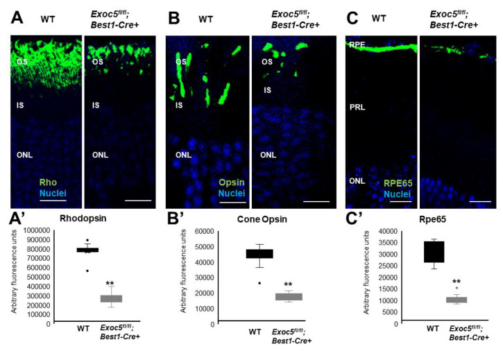
Figure 7 Immunohistochemical analysis of rod and cone photoreceptors in wild-type and conditional Exoc5 knockout mice at 27 weeks of age. Levels and localization of rhodopsin (green, Rho, (A)), Red/Green cone opsins (green, R/G Opsin, (B)), and RPE65 (green, RPE65, (C)), were assessed using immunohistochemistry in retinal sections from mice collected at 27 weeks of age to identify alterations in rod and cone outer segments and RPE. Rod and cone outer segments are dysmorphic, and RPE staining for RPE65 was severely disrupted. Image J was used to quantify rhodopsin (A’) and cone opsin (B’) fluorescence in photoreceptors, and RPE65 (C’) in RPE of WT and Exoc5fl/fl;Best1-Cre+ mice. Scale bars = 50 μm (A–C). OS, outer segments; IS, inner segments; ONL, outer nuclear layer, INL, inner nuclear layer; RPE, retinal pigmented epithelium. ** p < 0.05 (WT vs. Exoc5fl/fl;Best1-Cre+ mutants).