
|
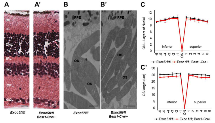
Figure 4 Histological and TEM analysis of retinas of Exoc5−/− mutants and WT mice at 20 weeks of age. (A,A’) H&E staining of WT and Exoc5−/− retinas at 20 weeks did not reveal histological differences. (B,B’) Transmission electron microscopy of wild-type (WT) mice photoreceptor cells show that rod OSs are shorter and thinner in Exoc5−/− retinas. The thickness of the ONL (C) and OS lengths (C’) from H&E sections through the optic nerve (ON; 0 μm distance from Optic Nerve and the starting point) was measured at 12 locations around the retina, six each in the superior and inferior hemispheres, each equally at 150 μm distances. (B,B’) Scale bars = 800 nm. OS, outer segments; RPE, retinal pigmented epithelium; IS, inner segments; ONL, outer nuclear layer; INL, inner nuclear layer; OPL, outer plexiform layer.