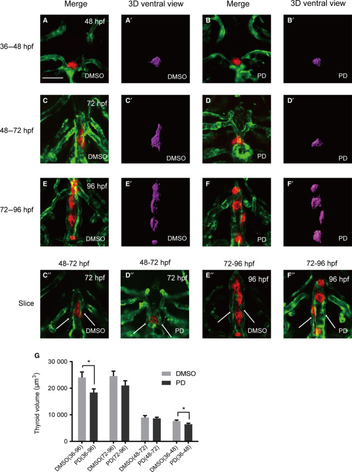
IMAGE

Fig. 4

ID
ZDB-IMAGE-210512-27
Source
Figures for Zhang et al., 2021
Image
Figure Caption

Fig. 4

Treatment of embryos with the FGFR1‐selective inhibitor PD166866 from 36 to 96 hpf caused severe defects in thyroid morphology and volume and in HAs. All embryos shown are oriented anterior to the top in the ventral views. (A, C, E) Analysis of DMSO‐treated embryos showed normal vascular development with normal thyroid morphology. (B) Treatment with PD 166866 from 36 to 48 hpf caused a reduction in the signal intensity of tg. (D) During 48 to 72 hpf drug treatments, the typical inverted ‘Y’ shapes of the thyroid were barely observed in embryos after drug treatment. (F) In response to drug treatment from 72 to 96 hpf, the thyroids possessed loose and short morphology compared with those in the nondrug treatment group according to 3D simulated images, and no obvious abnormal pharyngeal vasculature surrounding the thyroid was observed. (C’’‐F’’) To better visualize HA development in embryos, the slices of C to F were presented separately. (D’’) Abnormal HAs (stubby and malformed) were observed in all embryos during 48–72 hpf drug treatment. (G) Treatment with PD166866 from 36 to 48 hpf resulted in a reduction in thyroid volume compared with that observed in vehicle‐treated embryos (P < 0.05). Drug treatment from 36 to 96 hpf resulted in a significant reduction in thyroid volume (P < 0.05). Drug treatment from 48 to 72 hpf and 72 to 96 hpf resulted in a mild effect on thyroid volume. Arrow: HA. Scale bar: 50 μm. The error bars represent SEM. Data are representative of three independent experiments with similar results. We conducted statistical comparisons using Student's t‐test for quantitative measures. *P < 0.05.

Acknowledgments
This image is the copyrighted work of the attributed author or publisher, and ZFIN has permission only to display this image to its users. Additional permissions should be obtained from the applicable author or publisher of the image. Full text @ FEBS Open Bio