IMAGE

Figure 1

ID
ZDB-IMAGE-210510-3
Source
Figures for Bragazzi Cunha et al., 2021
Image
Figure Caption

Figure 1

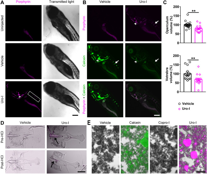
Zebrafish model of CEP develops bone phenotype resembling human disease. (A) 6dpf zebrafish larvae were injected with uro-I or vehicle and imaged by confocal microscopy at 7dpf. Porphyrin was detected only in the bones of uro-I-injected group. Arrowhead-operculum; box-vertebrae. (B) Larvae were treated as in (A) and injected with calcein prior to imaging. Arrowhead-operculum; arrow-4th vertebra. (C) Quantification of bone volume in larvae from (B); bone volume was normalized to vehicle-injected larvae set to 100%. Symbols represent individual larvae (14–18/group) from 4–5 independent experiments. (D) Larvae were treated as in (A). At 7dpf bones were harvested and imaged by epifluorescence microscopy pre and post HCl bone demineralization, arrow-notochord. (E) Hydroxyapatite was incubated with calcein/uro-I/copro-I and imaged by epifluorescence microscopy. Scale bars: 200 µm (A-D); 50 µm (E). Three-dimensional image reconstruction (A, B) was performed using Imaris 3D software v7.7 (http://imaris.oxinst.com/). **p < 0.01.

Figure Data
Acknowledgments
This image is the copyrighted work of the attributed author or publisher, and ZFIN has permission only to display this image to its users. Additional permissions should be obtained from the applicable author or publisher of the image. Full text @ Sci. Rep.