
|
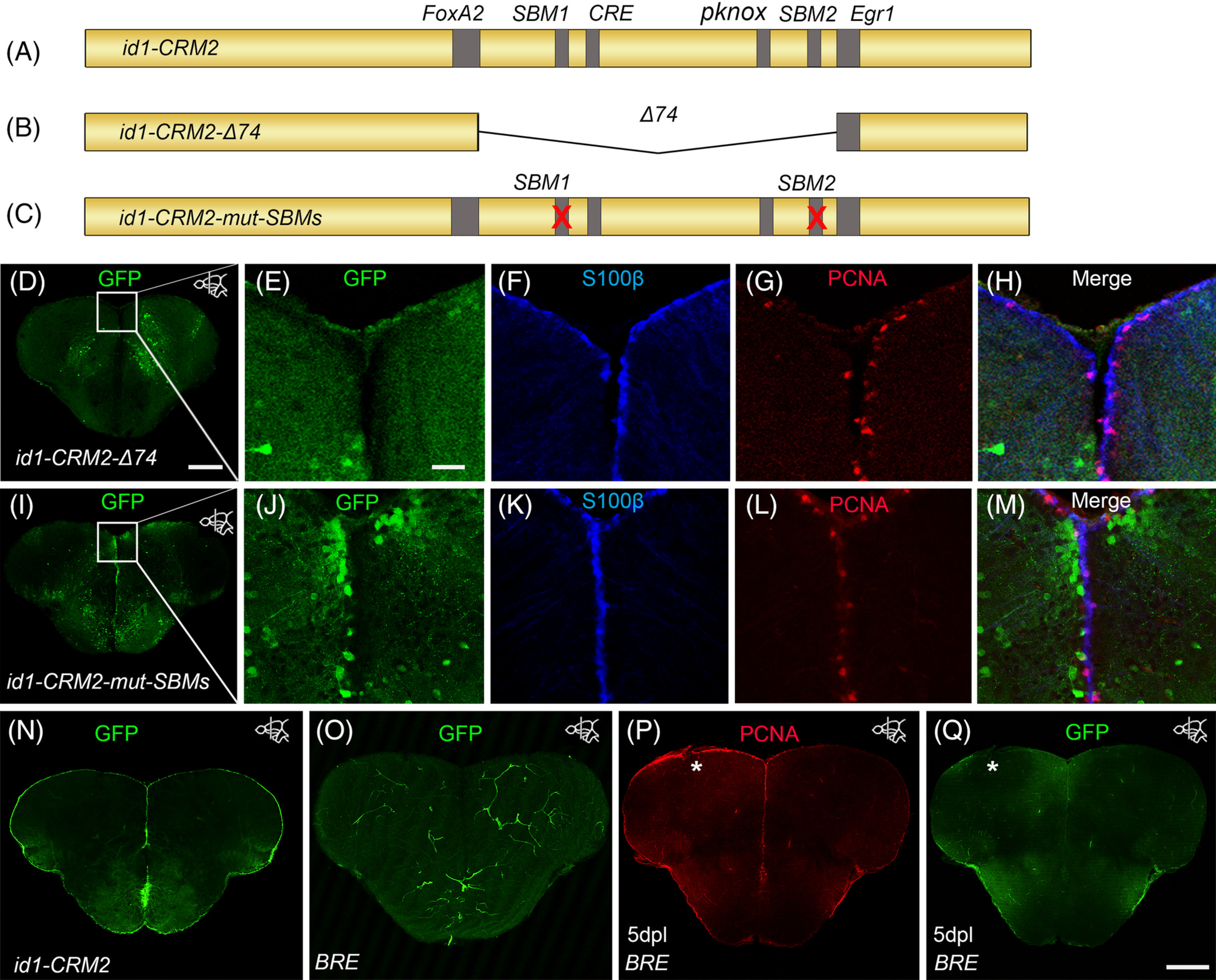
Fig. 4 The conserved BMP response element in the id1‐CRM2 is crucial for correct expression of the GFP reporter in the ventricular zone and RGCs. A‐C, Scheme showing mutated id1‐CRM2 reporter constructs: A, id1‐CRM2 wt construct with putative TF binding sites indicated in gray. B, id1‐CRM2‐Δ74 construct which contains a deletion of a 74 bp stretch of the most conserved sequence in id1‐CRM2. C, id1‐CRM2‐mut‐SBMs construct with mutations in the 2 SBMs (1 and 2) of id1‐CRM2. D, Deletion of the 74 bp stretch in the id1‐CRM2 abolished GFP expression in the ventricular zone. E‐H, Enlarged micrographs of D. E‐H, Immunohistochemistry with GFP (E), S100ß (F), and PCNA (G) antibodies on telencephalic cross sections of the id1‐CRM2‐Δ74 transgenic line shows no GFP reporter expression in S100ß+ RGCs (H, merged view). I‐M, Mutations in Smad binding motifs (SBM1 and 2) abolished GFP expression in the ventricular zone. J‐M, Magnification of white‐boxed region in I. J‐M, Immunohistochemistry with GFP (J), S100ß (K), and PCNA (L) antibodies on telencephalic cross sections of the id1‐CRM2‐mut‐SBMs transgenic line show no colocalization between the RGC marker, S100ß, and GFP (M, merged view). N, GFP expression driven by id1‐CRM2:GFP reporter construct in the ventricular zone (control). O, P, Q Immunohistochemistry of telencephalic cross sections with GFP (O, Q) and PCNA (P). O, Q, The BRE does not drive GFP expression in the RGCs (O) and is not inducible by telencephalic injury (Q). The left injured side is labeled with a white asterisk. Anteroposterior positions of transverse sections are indicated in the upper right‐hand corner of each image. Scale bar = 20 μm (E, F, G, H, J, K, L, M); 200 μm (D, I, N, O, P, Q)