
Image
|
Figure Caption
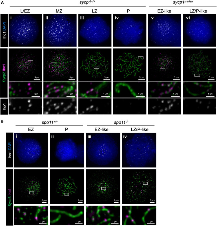
FIGURE 8 Iho1 localization in sycp1 and spo11 mutant spermatocytes. (A) costaining of Iho1 and Sycp2 in sycp1+/+ (i to iv) and sycp1isa/isa (v and vi) spermatocytes. Regions marked in white rectangles are shown at a higher magnification at the bottom. (B) Costaining of Iho1 and Sycp3 in spo11+/+ (i and ii) and spo11–/– (iii and iv) spermatocytes. Regions marked in white rectangles are shown at a higher magnification at the bottom.
Acknowledgments
This image is the copyrighted work of the attributed author or publisher, and
ZFIN has permission only to display this image to its users.
Additional permissions should be obtained from the applicable author or publisher of the image.
Full text @ Front Cell Dev Biol