Image
Figure Caption

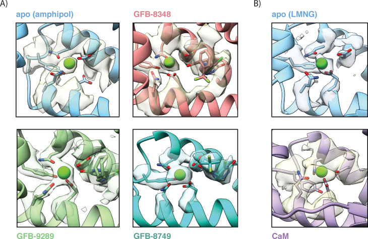
Figure 4—figure supplement 2. Ca<sup>2+</sup>-binding site in the VSL domain of apo and ligand-bound TRPC4.

(A) Close-up view of the Ca2+-binding site in apo and ligand-bound TRPC4 determined in amphipols. The coordinating residues in the Ca2+ ion binding site and the modelled Ca2+ ion are shown in stick and sphere representation along with their densities. The oxygen atom of the ligand molecules is situated close to the bound Ca2+-ion (refer)Figure 4C (B) Close-up view of the Ca2+-binding site in apo and TRPC4-CaM complex determined in presence of lauryl maltose neopentyl glycol (LMNG). The external addition of Ca2+ ion during the TRPC4-CaM complex preparation reflects its strong density. The different structural forms shown in (A) and (B) are color coded and named accordingly.

Acknowledgments
This image is the copyrighted work of the attributed author or publisher, and ZFIN has permission only to display this image to its users. Additional permissions should be obtained from the applicable author or publisher of the image. Full text @ Elife