
|
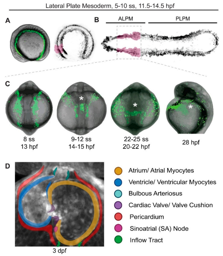
Figure 1 Earliest stages of cardiac development in zebrafish. Stages of zebrafish heart formation from lateral plate mesoderm (LPM). (A,B) LPM during early somitogenesis on three-dimensional (A) and virtually flat-mounted (B) zebrafish embryo, anterior to the left. The prospective bilateral heart field is marked in magenta within the anterior LPM (ALPM). (C) Stages of heart field convergence at the midline, formation of the cardiac disc and cone, migration to the left, and early heart tube stages; maximal projection of z-stack of drl:EGFP transgenics [24], anterior (head) to the top, with the embryo curved over the yolk at this stage (tail end behind the yolk in the first three panels, out of view in the right-most panel). Note how the bilateral ALPM migrates to the midline and forms the leftward-moving heart tube (asterisks). (D) 72 hpf (3 dpf) zebrafish heart with all major parts color-coded, anterior/rostral to the top; greyscale max projection of z-stack of ubi:Zebrabow transgenics overlaid with annotation of individual structures [25]. See text for details, images not to scale.