
Figure 2
- ID
- ZDB-IMAGE-210210-30
- Publication
- Kuil et al., 2021 - Zebrafish: A Model Organism for Studying Enteric Nervous System Development and Disease
- All Figures
- Figures for Kuil et al., 2021
|
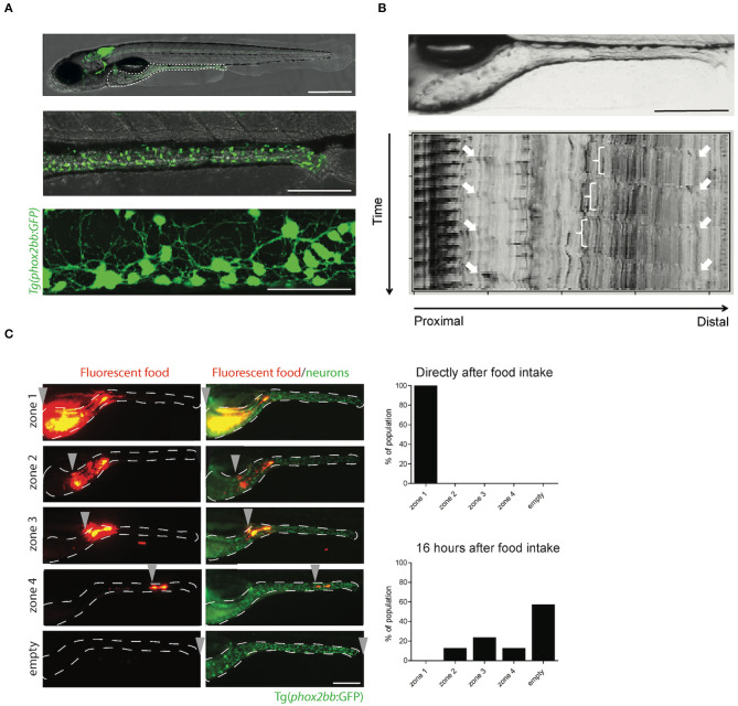
Figure 2
The transparency of zebrafish larvae allows non-invasive visualization of enteric neurons