IMAGE

Fig 2

ID
ZDB-IMAGE-210204-35
Source
Figures for Yergert et al., 2021
Image
Figure Caption

Fig 2 Endogenous <italic>mbpa</italic> mRNA localizes to myelin sheaths between 3–5 dpf.

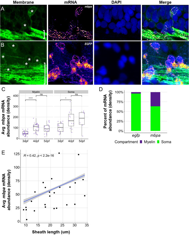
(A and B) Representative images of smFISH experiments using 4 dpf transgenic larva expressing EGFP-CAAX to mark oligodendrocytes. Images show sagittal sections of the hindbrain. DAPI stain labels nuclei. Sections were treated with smFISH probes designed to detect mbpa (A) or egfp (B) mRNA. Asterisks mark cell bodies and brackets mark myelin tracts. Scale bars, 10 μm. (C) Average mbpa mRNA density per cell body or equivalent volume of myelin from 3 to 5 dpf. Density was measured using the integrated density of fluorescence intensity in cell bodies and approximately equal volumes of myelin along the myelin tracts. A minimum (n) for each group was 3 larvae, 6 cell bodies, and 15 myelin regions. Statistical significance evaluated using Wilcoxon test. (D) Proportion of egfp or mbpa mRNA abundance in cell bodies compared to myelin tracts. A minimum (n) for each group was 3 larvae, 11 cell bodies, and 21 myelin regions. (E) Average mbpa mRNA density within individual sheaths plotted as a function of sheath length. Statistical significance evaluated using Spearman’s correlation coefficient. Shaded area represents 95% confidence interval. n = 7 embryos, 26 sheaths. The underlying numerical data can be found in S5S7 Data. dpf, days post fertilization; smFISH, single molecule fluorescent in situ hybridization.

Acknowledgments
This image is the copyrighted work of the attributed author or publisher, and ZFIN has permission only to display this image to its users. Additional permissions should be obtained from the applicable author or publisher of the image. Full text @ PLoS Biol.