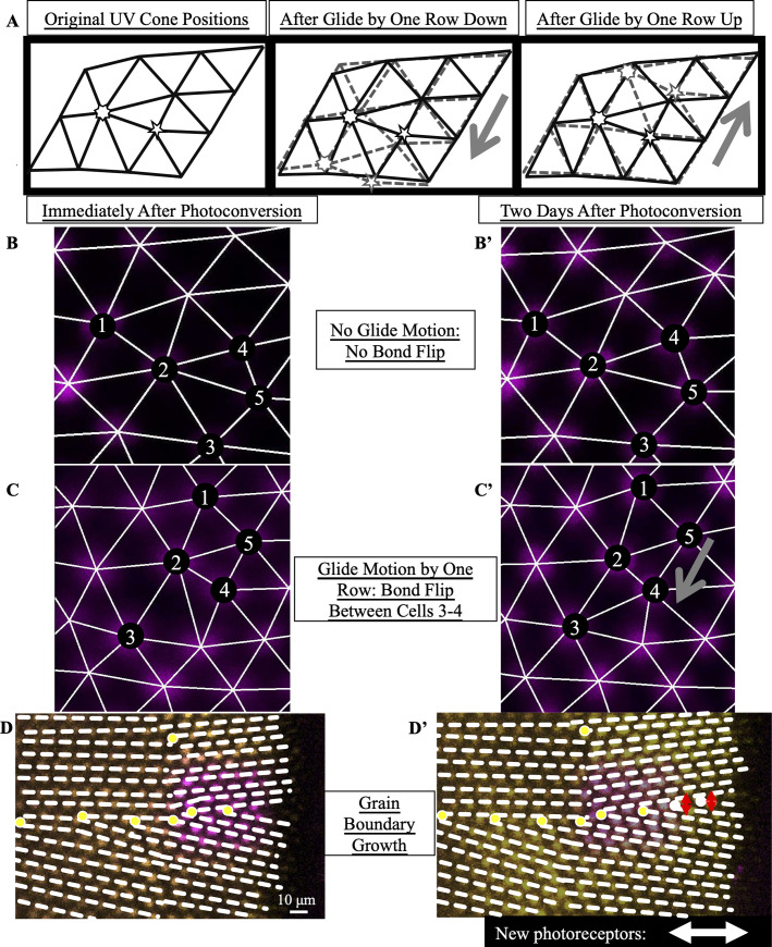
IMAGE

Fig 5

ID
ZDB-IMAGE-210113-122
Source
Figures for Nunley et al., 2020
Image
Figure Caption

Fig 5 By estimating the timescale of Y-Junction motion, we conclude that Y-Junctions line up into grain boundaries during initial mosaic formation rather than by subsequent Y-Junction motion.

(A) Expected motion of individual UV cones in the case of glide motion by one row in either direction. Left triangulation shows UV cones near the defect core; a UV cone sits on each site of triangulation. The center and right panels overlay the positions of UV cones before (black) and after (gray) glide in the direction denoted by the gray arrows. Note that the originally five- and seven-coordinated UV cones in the black triangulation both become six-coordinated. (B,B’) Example of Y-Junction in photoconverted region in which no bond flips in two days. The photoconverted fluorescent signal in UV cone nuclei is pseudo-colored magenta. For reference, the same five cones are numbered in both images. White lines: triangulation of UV cones. (C,C’) Y-Junction in photoconverted region from Fig 4B. One bond has flipped in the triangulation over two days, meaning that the Y-Junction has glided in the direction of the gray arrow by a row. (D,D’) Observation of grain boundary growth during initial mosaic formation. Immediately after photoconversion, one observes seven Y-Junctions (yellow dots), six within a grain boundary and an isolated Y-Junction nearby. White dashed lines: rows of UV cones. Two days later, one observes two additional Y-Junctions in the grain boundary. Based on the constraint that a Y-Junction does not glide faster than one row in two days, Y-Junctions must have initially formed within the regions indicated by red arrows (thus, within the grain boundary). White double-headed arrow: columns of cones incorporated since photoconversion.

Acknowledgments
This image is the copyrighted work of the attributed author or publisher, and ZFIN has permission only to display this image to its users. Additional permissions should be obtained from the applicable author or publisher of the image. Full text @ PLoS Comput. Biol.