IMAGE

Figure 2

ID
ZDB-IMAGE-201121-58
Source
Figures for Rodrigues da Silva et al., 2020
Image
Figure Caption

Figure 2

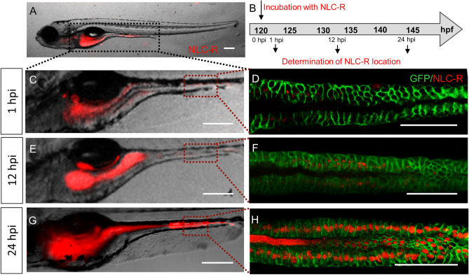
Absorption of NLCs in the larval body. (A) TgBAC(cldn15la:GFP)pd1034 larvae, at 5 days post fertilization (dpf), exposed to NLCs loaded with rhodamine-PE, by the immersion method. The larvae intestine is demarked with a black dotted line. (B) Timeline: fluorescence was monitored at 1, 12 and 24-h post incubation (hpi). Representative images of larvae examined under a fluorescence stereomicroscope at 1 hpi (C), 12 hpi (E) and 24 hpi (G). A region of the mid-intestine of TgBAC(cldn15la:GFP)pd1034 larvae, demarked by a red dotted line in (C,E,G) was examined under confocal microscope at 1, 12 and 24 hpi (D,F,H, respectively). NLC-R: NLCs labeled with red fluorescent rhodamine-PE. Scale bar 200 μm. The images in A, CH were edited with ImageJ software v.1.52a (https://imagej.nih.gov/ij/).

Acknowledgments
This image is the copyrighted work of the attributed author or publisher, and ZFIN has permission only to display this image to its users. Additional permissions should be obtained from the applicable author or publisher of the image. Full text @ Sci. Rep.