
|
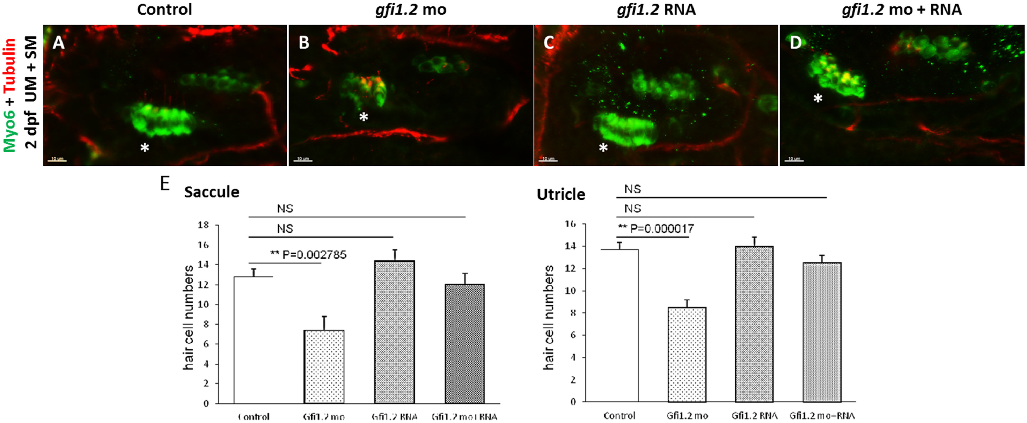
Fig. 4 The number of hair cells and neurons was significantly decreased by MO-mediated gfi1.2 knockdown and can be rescued by exogenous gfi1.2. Neurons were labeled by the immunostaining of Myo6 and tubulin at 2 dpf. The number of neurons in the zebrafish neuromast was compared in (A) wild-type (wt), (B) MO-mediated gfi1.2-knockdown, (C) gfi1.2 mRNA-overexpressing, and (D) MO gfi1.2 + gfi1.2 mRNA. Hair cells were also labeled by the immunostaining of Myo6 and tubulin at 2 dpf. The number of hair cells in the zebrafish inner ear was compared in (E) wild-type (wt), (F) MO-mediated gfi1.2-knockdown, (G) gfi1.2 mRNA-overexpressing, and (H) MO gfi1.2 + gfi1.2 mRNA. In the gfi1.2 mutants, hair cells in the utricle maculae (UM) and saccule maculae (SM) significantly decreased in number (p < 0.05). Over-expression of gfi1.2 in the wild-type zebrafish did not affect the number of hair cell number (E). NS: not significant. The utricle is indicated by a white *, and the contralateral structure is the saccule.
Reprinted from Hearing Research, 396, Yu, R., Wang, P., Chen, X.W., The role of gfi1.2 in the development of zebrafish inner ear, 108055, Copyright (2020) with permission from Elsevier. Full text @ Hear. Res.