
|
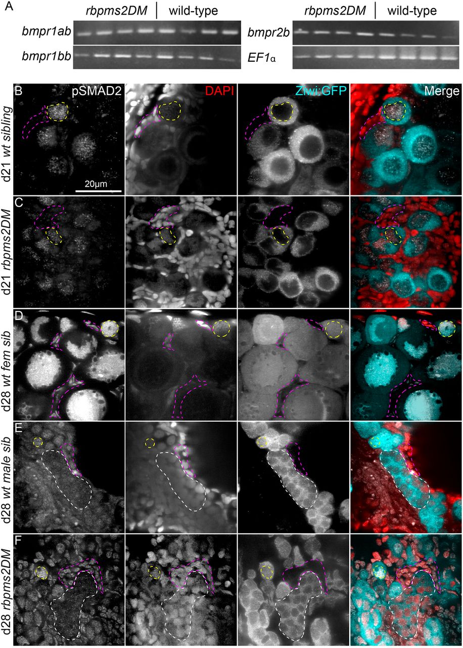
Fig. 1 Molecularly bipotential juvenile gonads of rbpms2DMs and activation of p-SMAD in early germ cells and differentiating gonads. (A) RT-PCR on dissected trunks from individual d21 rbpms2DMs and wild-type siblings reveals no differences in expression of relevant receptors. (B-F) p-Smad2, DAPI and ziwi:GFP staining. Representative slices from Z-series reveal localization of p-Smad2 in bipotential and early differentiating gonads of d21 and d28 juvenile zebrafish, respectively. In undifferentiated bipotential ovaries of both wild-type (n=3) (B) and rbpms2DM (n=3) (C), p-Smad2 is detected in the nuclei of germ cells (example outlined with yellow dashed line) but not the nuclei of the surrounding somatic cells (examples outlined in pink dashed lines). (D) By d28 in early differentiated females (n=4), p-Smad2 remained localized to the germ cell nuclei and began to be expressed in somatic nuclei. Similarly, p-Smad2 was expressed at d28 in both germ cell nuclei (examples outlined in white dashed lines) and somatic nuclei of wild-type males (n=4) (E) and rbpms2DM gonads (n=4) (F). ‘Wild-type’ denotes wild-type or heterozygous animals as well as rbpms2a or rbpms2b single mutants that are heterozygous or homozygous for the wild-type allele at the other rbpms2 locus because these animals are phenotypically normal.