
Image
|
Figure Caption
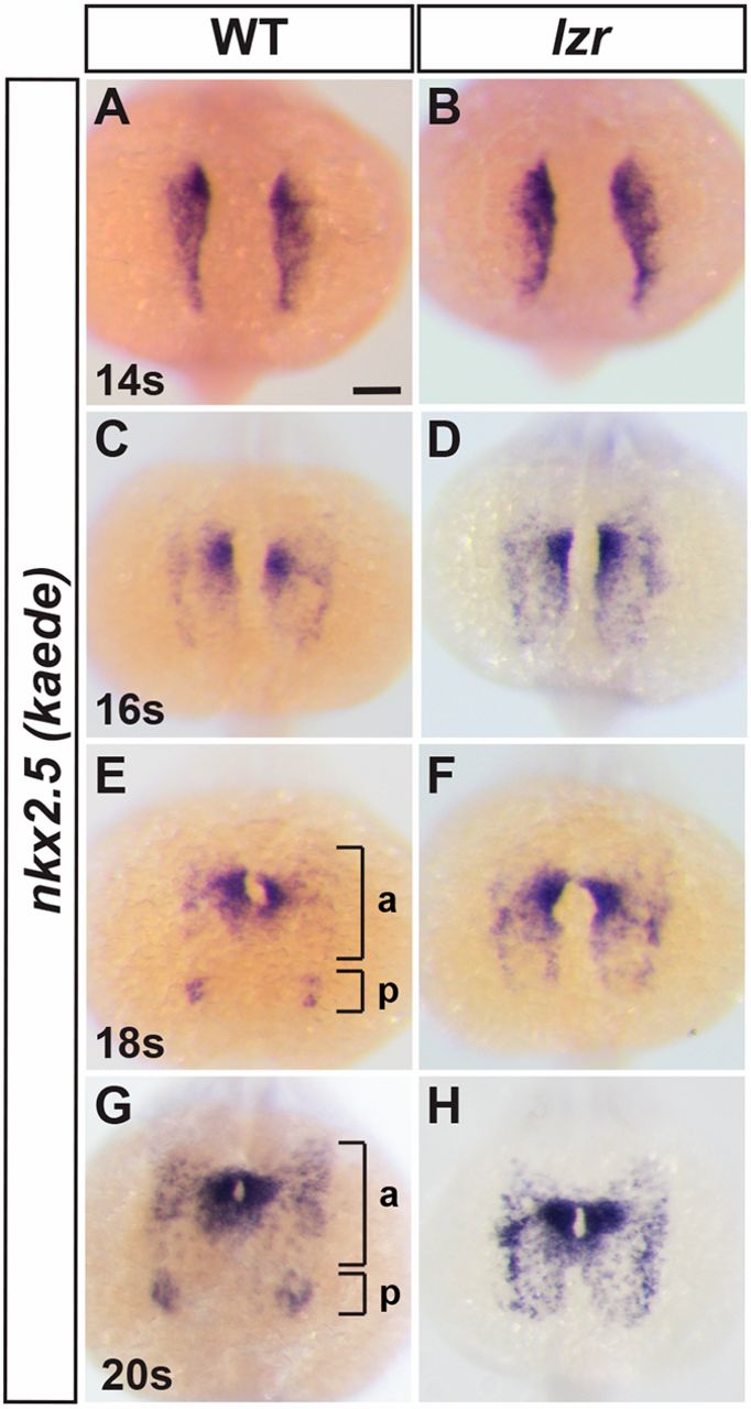
Fig. 7 Nkx2.5+ progenitors fail to segregate into cardiac and endothelial populations in lzr mutants. (A-H) ISH for nkx2.5:Kaede in WT and lzr embryos at 14 s, 16 s, 18 s and 20 s. Views are dorsal with anterior up. Brackets in E and G denote anterior (a) and posterior (p) nkx2.5:Kaede+ clusters. 14 s: WT (n=47), lzr (n=5); 16 s: WT (n=26), lzr (n=6); 18 s: WT (n=29), lzr (n=6); 20 s: WT (n=58), lzr (n=6). Scale bar: 100 µm.
Figure Data
Acknowledgments
This image is the copyrighted work of the attributed author or publisher, and
ZFIN has permission only to display this image to its users.
Additional permissions should be obtained from the applicable author or publisher of the image.
Full text @ Development