IMAGE

Figure 7

ID
ZDB-IMAGE-200413-23
Source
Figures for Sie et al., 2020
Image
Figure Caption

Figure 7

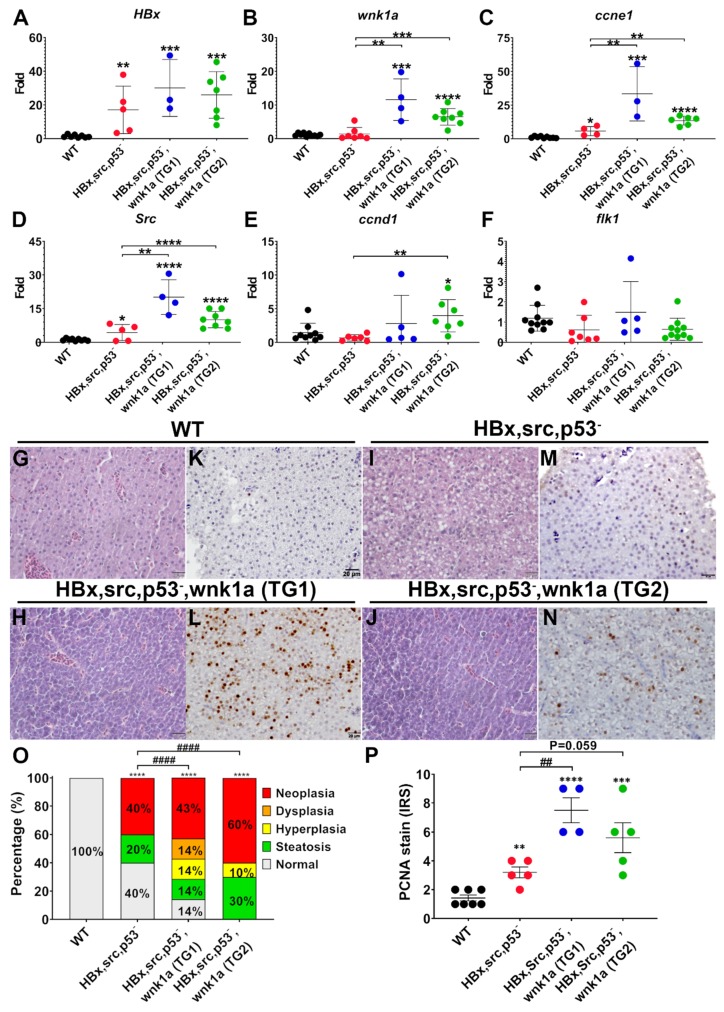
Endothelial cell-specific wnk1a expression promote tumorigenesis in HCC-transgenic fish. (AF) QPCR analysis of genes in WT, HBx, src(p53-) fish, and HBx, src(p53-) fish overexpressing wnk1a in endothelium. Two separate transgenic lines, TG1 and TG2, were studied. (GJ) Representative images of H&E-stained liver sections. Scale bar = 20 μm. (K–N) Representative images of immunohistochemical staining of liver sections for proliferating cell nuclear antigen (PCNA). Scale bar = 20 μm. The statistical significance was analyzed by unpaired Student’s t-test. **: 0.001 < p ≤ 0.01; ***: 0.0001 < p ≤ 0.001; ****: p ≤ 0.0001 compared to WT control. (O) Distribution of histopathological findings of H&E-stained liver sections as shown in panels (G–J). The statistical significance was analyzed by Chi-square test. ****: p ≤ 0.0001 compared to WT control, ####: p ≤0.0001 comparison between transgenic fish. (P) Average immunoreactive score (IRS) of proliferating cell nuclear antigen (PCNA) staining as shown in panels K-N. **: 0.001 < p ≤ 0.01; ***: 0.0001 < p ≤ 0.001; ****: p ≤ 0.0001 compared to WT control. ##: 0.001 < p ≤ 0.01 comparison between transgenic fish.

Acknowledgments
This image is the copyrighted work of the attributed author or publisher, and ZFIN has permission only to display this image to its users. Additional permissions should be obtained from the applicable author or publisher of the image. Full text @ Cancers