IMAGE

Figure 2

ID
ZDB-IMAGE-200413-18
Source
Figures for Sie et al., 2020
Image
Figure Caption

Figure 2

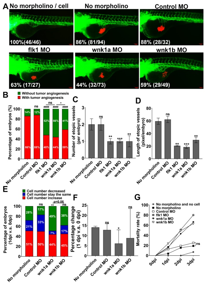
Effect of wnk1 knockdown on tumor-induced angiogenesis and tumor cell proliferation in zebrafish embryos. (A) Representative images of control embryos and embryos injected with Hep3B_Lifeact-RFP ± indicated morpholino. Images were at 2 dpi. Percentage of live embryos (number of live embryos/total embryos) for each experimental group was shown. Scale bar = 100 μm. (B) Percentage of embryos with (red zone) and without angiogenic response (green zone). Total number of embryos was 24. The statistical significance was analyzed by Chi-square test. ####: p ≤ 0.0001; ns: non-significant compared to the no morpholino control. ns: non-significant compared flk1 MO and wnk1a MO; *: 0.01 < p ≤ 0.05 compared wnk1a MO and wnk1b MO. (C,D) The average number of ectopic vessels (C) and length of ectopic vessels (D) in different morphants. The statistical significance was analyzed by unpaired Student’s t-test. **: 0.001 < p ≤ 0.01; ***: 0.0001 < p ≤ 0.001 compared to the no morpholino control. (E) Distribution of embryos with decreases (green zone), unchanged (blue) and increases in tumor-associated fluorescence in 1 dpi relative to 0 dpi. Changes of more than 5% on either direction were defined as decrease or increase. Total number of embryos was 24. The statistical significance was analyzed by Chi-square test. **: 0.001 < p ≤ 0.01; ns: non-significant compared to the no morpholino control. (F) Percentage changes of the tumor-associated fluorescent intensity in 1 dpi from 0 dpi. ns: non-significant, p > 0.05; *: 0.01 < p ≤ 0.05 compared to the no morpholino control. (G) Mortality rate of different morphants.

Acknowledgments
This image is the copyrighted work of the attributed author or publisher, and ZFIN has permission only to display this image to its users. Additional permissions should be obtained from the applicable author or publisher of the image. Full text @ Cancers