
|
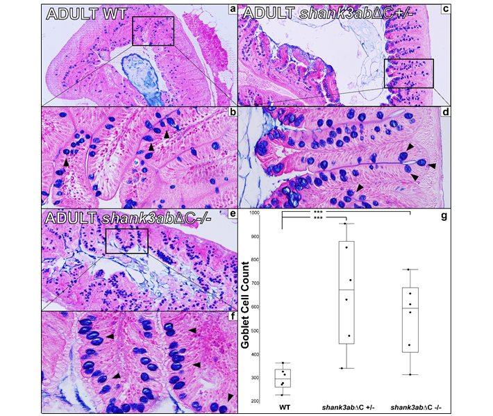
Fig. S2 Figure S2 a Transverse 5μm section of adult WT upper intestinal tissue stained with alcian blue and Eosin B (n=6 for WT, n=6 for shank3abΔC +/- and n=6 for shank3abΔC -/-). b 40x magnification shows dense plicae that extend to the point of nearly occluding the luminal space. c,d In shank3abΔC +/- upper intestinal tissue, increased counts of goblet cells (black arrowheads) suggest inflammation. e,f Homozygous shank3abΔC -/- adults also show increased goblet cell count. Comparison of goblet cell counts in WT, shank3abΔC +/-, and shank3abΔC -/- adults show a significant increase in the number of goblet cells in both shank3abΔC +/- and shank3abΔC -/- adults. WT and shank3abΔC -/- comparison (p<0.0138) and WT and shank3abΔC +/- comparison (p<0.0015).