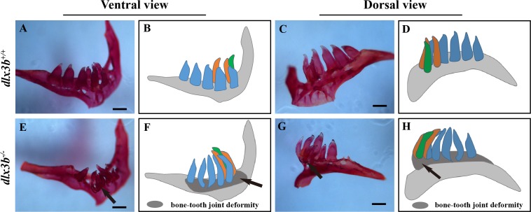
IMAGE

Figure 4

ID
ZDB-IMAGE-200306-29
Source
Figures for Pang et al., 2020
Image
Figure Caption

Figure 4 Pharyngeal teeth of homozygous mutants (<italic>dlx3b</italic><sup>−∕−</sup>) exhibited deformiy.

Compared with the pharyngeal teeth of the wild-type (A, C), those of the mutant group exhibited abnormal bending towards the dorsal side (E, G) with the abnormal bone growth of the pharyngeal bone at the bone-tooth joint (E, G, black arrow). (A–D) The pharyngeal dentition of the wild-type sibling; (E–H) the pharyngeal dentition of the dlx3b−∕− mutant. (A, E) The pharyngeal dentition viewed from the ventral side. (C, G) The pharyngeal dentition viewed from the dorsal side. (B, D, F, H) Schematic representations of A, C, E, and G, respectively (ventral tooth row, blue; mediodorsal tooth row, ochre; dorsal tooth row, green; replacement teeth not shown; and the abnormal bone growth is depicted as a dark gray region in F and H). Scale bar: 200 µm. n = 8 for each group.

Figure Data
Acknowledgments
This image is the copyrighted work of the attributed author or publisher, and ZFIN has permission only to display this image to its users. Additional permissions should be obtained from the applicable author or publisher of the image. Full text @ Peer J.