IMAGE

Figure 4

ID
ZDB-IMAGE-200306-20
Source
Figures for Scheib et al., 2020
Image
Figure Caption

Figure 4

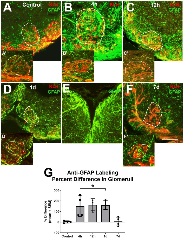
Higher magnification analysis of Z-stacks of anti-GFAP (green) and anti-keyhole limpet hemocyanin (KLH; red) labeling during repetitive damage to the olfactory organ. (A) Anti-KLH allowed identification of a ventral medial glomerulus (dashed outline). Anti-GFAP labeled processes within the glomerulus in the right olfactory bulb (dashed outline) varied in size and length and had no clear organization in untreated control bulbs. (A’) A glomerulus (dotted outline) in the same region of the left olfactory bulb of the same fish displayed similar morphology of anti-GFAP labeled processes, n = 4. (B) After 4 h of damage to the olfactory organ, anti-GFAP labeled processes appeared to be more numerous and thicker within the glomerulus (dashed outline) of the affected bulb when compared to the comparable glomerulus (dotted outline) of the internal control bulb (B’), n = 4. (C) By 12 h after damage to the periphery, there were numerous anti-GFAP labeled processes associated with the glomerulus (dashed outline) in the affected bulb that varied in length and thickness, while the glomerulus (dotted outline) in the internal control bulb (C’) retained typical labeling, n = 3. (D) At 1 day after repeated damage to the olfactory organ, affected glomeruli (dashed outline) still appeared to have thicker and more numerous anti-GFAP labeled processes when compared to glomeruli (dotted outline) in the internal control bulb (D’), n = 3. (E) Side by side comparison of both bulbs at 1 day showed that the left, internal control bulb retained control levels of anti-GFAP labeling, while the right, treated bulb showed more and thicker labeled processes. (F) By 7 days after repetitive peripheral damage, anti-GFAP labeled processes in glomeruli (dashed outline) in the affected bulb appeared to have similar morphology to anti-GFAP labeled processes in glomeruli (dotted outline) of the internal control bulb (F’), n = 4. Scale bar = 20 μm for all. (G) The percent difference of anti-GFAP optical density (OD) measurements between treated and untreated glomeruli was significantly different at 4 h, 12 h, and 1 day of repetitive peripheral damage but was no longer significantly different after 7 days of repeated damage. *p < 0.05.

Acknowledgments
This image is the copyrighted work of the attributed author or publisher, and ZFIN has permission only to display this image to its users. Additional permissions should be obtained from the applicable author or publisher of the image. Full text @ Front. Neuroanat.