
|
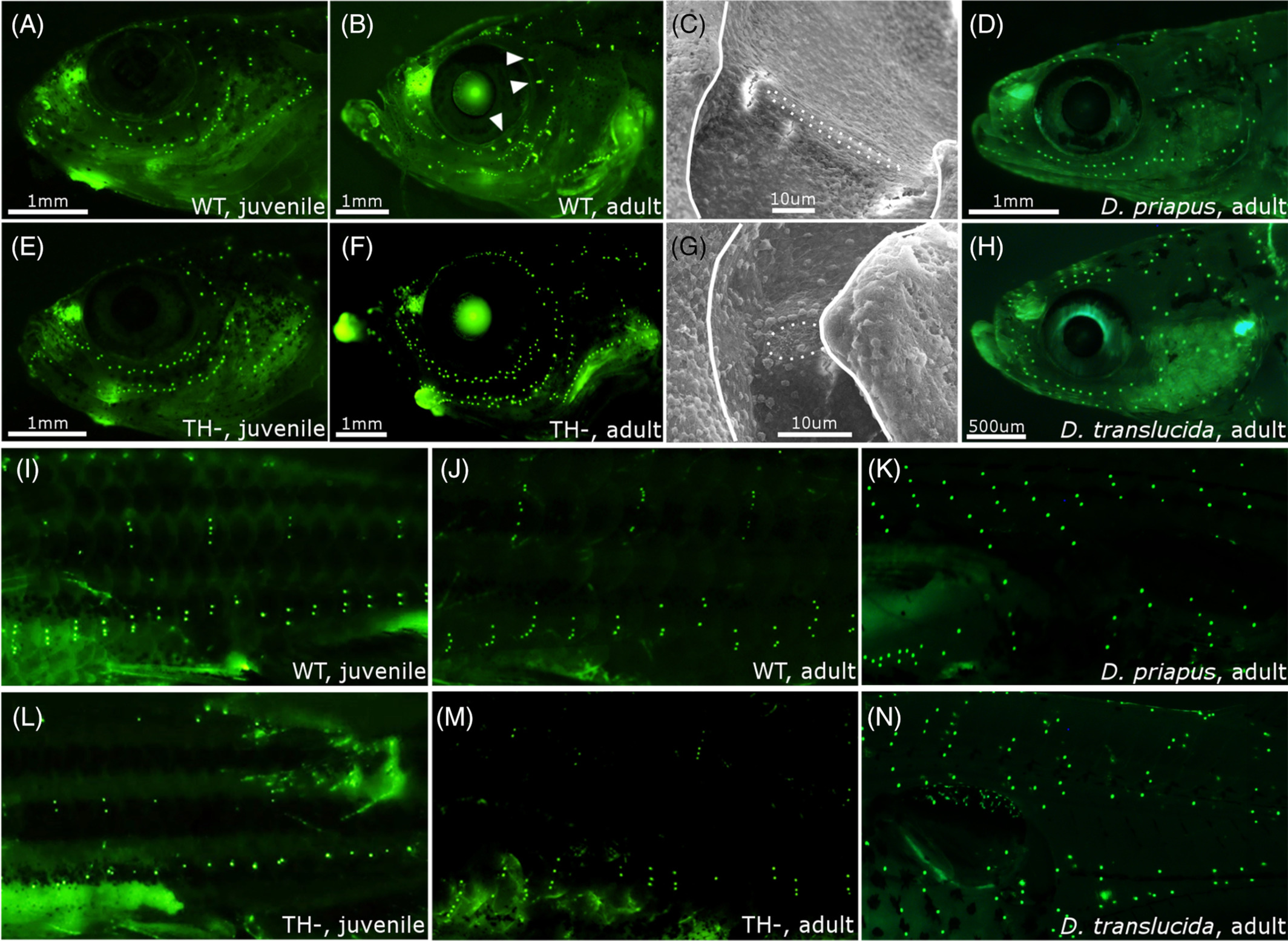
Fig. 4
The lateral line system in zebrafish and Danionella. A and B, DiASP live staining of head neuromasts in WT juvenile (13 mm SL) and adult (18+ mm SL). Arrowhead indicates rod‐shaped canal neuromasts at adult stage. C, Scanning electron micrographs of a supraorbital canal neuromast in WT adult. Dotted line outlines the neuromast; solid line outlines the bony edge of the canal. D, Head neuromasts in Danionella priapus adult. E‐G, Same as B‐D in TH− zebrafish. H, Facial neuromasts in Danionella translucida adult. I‐N, DiASP live staining of trunk neuromasts in WT juvenile zebrafish (13 mm SL, I), WT adult (18+ mm SL, J), D. priapus adult (K), TH− juvenile (13 mm SL, L) TH− adult (18+ mm SL, M), and D. translucida adult (N). SL, standard length; TH−, hypothyroid; WT, Wild‐type