
|
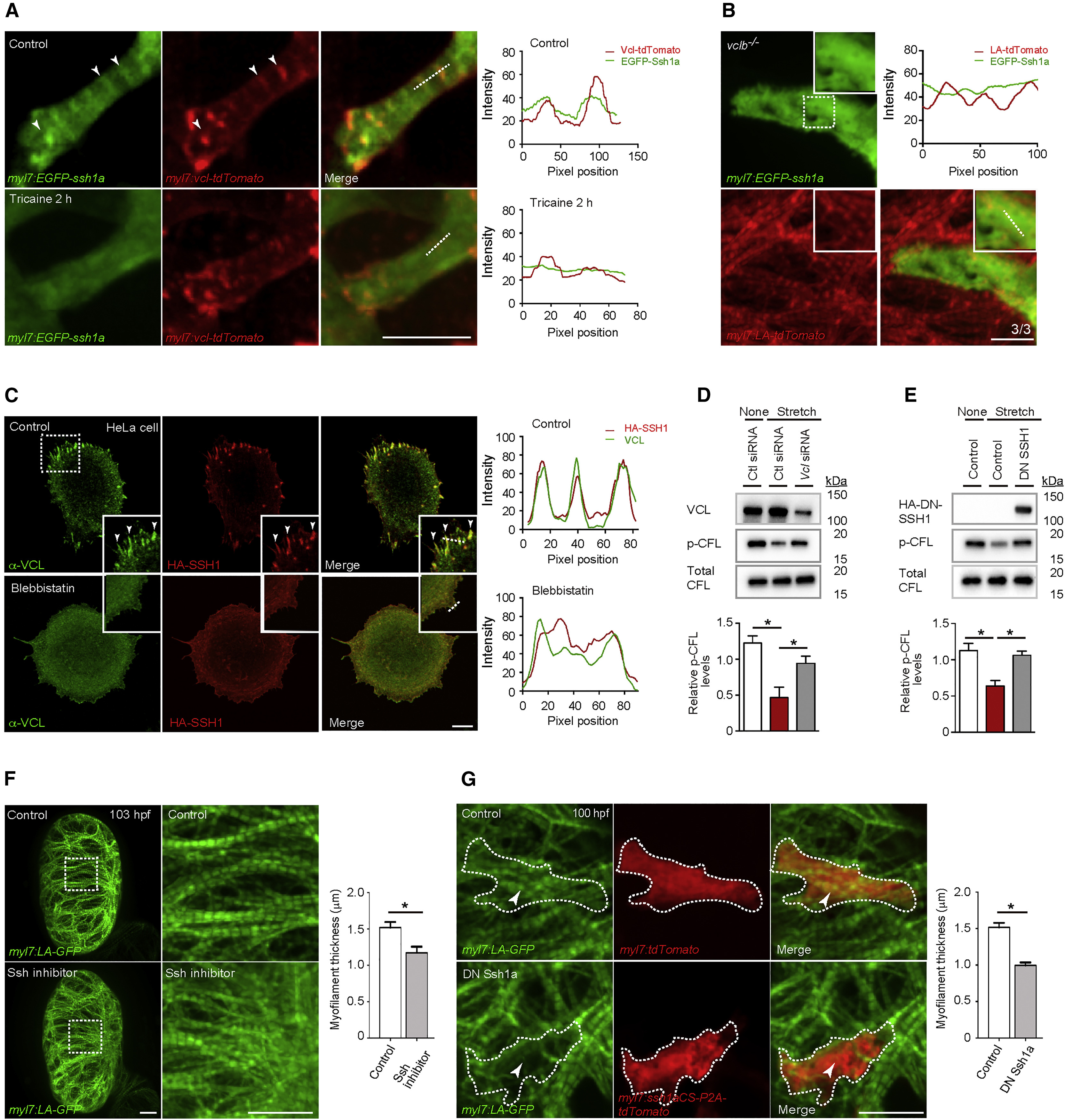
Fig. 6
SSH1 Regulates Cardiomyocyte Myofilament Maturation
(A) 3D images of a 103 hpf EGFP-Ssh1a expressing ventricle immediately after cardiac arrest and 2 h after tricaine treatment; fluorescence intensities along white dashed lines. EGFP-Ssh1a colocalizes with Vcl-tdTomato in CMs (arrowheads), but this co-localization is abrogated by tricaine treatment.
(B) 3D images of a 103 hpf vclb−/− ventricle expressing EGFP-Ssh1a; fluorescence intensities along white dashed line (n = 3 animals; each animal contains 2–4 EGFP-Ssh1a+ CMs).
(C) VCL and HA immunostaining of HeLa cells treated without or with blebbistatin; fluorescence intensities along white dashed lines. HA-SSH1 colocalizes with VCL (arrowheads), and blebbistatin treatment leads to loss of this co-localization.
(D) Protein expression after siRNA transfection in mouse neonatal CMs exposed to cyclic stretch; relative levels of p-CFL (n = 4).
(E) Protein expression in RNCMs exposed to cyclic stretch; relative levels of p-CFL (n = 3).
(F) 3D images of 103 hpf hearts treated without or with Ssh inhibitor from 72 to 103 hpf. Myofilament thickness measured in 103 hpf ventricles (n = 5 ventricles).
(G) 3D images of 100 hpf ventricle. DN Ssh1a overexpression leads to myofilament disruption (arrowheads). Myofilament thickness measured in 100 hpf CMs (n = 7–10 CMs). Error bars, SEM. ∗p < 0.05 by ANOVA followed by Tukey’s HSD test. Scale bars, 20 μm in (A), (B), (F), and (G); 10 μm in (C).
Reprinted from Developmental Cell, 51(1), Fukuda, R., Gunawan, F., Ramadass, R., Beisaw, A., Konzer, A., Mullapudi, S.T., Gentile, A., Maischein, H.M., Graumann, J., Stainier, D.Y.R., Mechanical Forces Regulate Cardiomyocyte Myofilament Maturation via the VCL-SSH1-CFL Axis, 62-77.e5, Copyright (2019) with permission from Elsevier. Full text @ Dev. Cell